Fig. 12.1
Supine position. The neck is placed in neutral position. The arms are abducted <90°, supinated, and padded underneath. All other pressure points are padded with a cushioned mattress
The lumbosacral area and occiput should also be provided padding underneath for prevention of pressure sores. In addition, it is imperative to periodically rotate the head during long or extended procedures to redistribute the weight and evade pressure alopecia secondary to hair follicle ischemia [7]. Patients with a history of chronic back pain or kyphoscoliosis may require additional padding or even slight flexion of the bed at the hip to avoid an exacerbation of their condition. Any bony prominences in contact with the operating table should also be padded, including the heels. Caution should also be used to prevent pressure or leaning on the lower extremities by the operating room (OR) staff which can inhibit venous return and increase the risk of deep venous thrombosis.
Additionally, placing the patient in the frog leg position while supine, with the hips and knees flexed and externally rotated, can facilitate access to the perineum, genitalia, and rectum if indicated and is commonly used during urinary catheter placement.
Physiologic Changes Associated with the Supine Position
Cardiovascular
The supine position benefits from having the least hemodynamic or ventilatory changes associated with it. Transferring from the erect to supine position facilitates venous drainage from the lower extremities, increasing right sided filling pressures, thereby increasing cardiac output. Minimal changes, however, are seen in blood pressure due to a reflex decrease in heart rate from the improved venous return. Given that the patient remains level on the operating table, equal pressures throughout the arterial system are appreciated. Caution is warranted though, in certain conditions that may increase intra-abdominal pressure such as tumor, obesity, ascites, or a gravid uterus, as they may impede venous return in the supine position, leading to hypotension.
Respiratory
Patient positioning and the effects of anesthesia significantly alter lung volumes and mechanics. The functional residual capacity (FRC), which is the sum of the residual and expiratory reserve volumes, is of particular importance to the anesthesiologist. The FRC is the volume of lung remaining after a normal tidal volume exhalation. A change in position from standing to supine reduces FRC by approximately 800 mL in the average adult [8]. Induction of anesthesia with muscle relaxation further decreases FRC by 20% [9]. When FRC is reduced, there is a greater likelihood that it will drop below the closing capacity (i.e., the volume at which the physical forces promoting small airway closure overcome the inherent elasticity of the lung tissue). This effect leads to atelectasis, pulmonary shunting, and a reduction in PaO2.
While supine, gravity assists in increasing perfusion to the dependent, or posterior, lung segments, which is favored during spontaneous breathing; however, during controlled ventilation, the independent anterior segments are favored, leading to an increase in ventilation/perfusion mismatch.
Potential Complications (Table 12.1)
Peripheral Nerves (Ulnar, Radial, and Median)
Ulnar neuropathy is the most frequent site (28%) of anesthesia-related nerve injury according to the ASA Closed Claims Database [10]. The ulnar nerve is susceptible to excessive pressure as it courses through the medial elbow in the post-condylar groove [11]. Proper padding should be applied to prevent extended periods of pressure in this area, and rotating the arm into a supinated or neutral position should also prevent its occurrence [12]. When the forearm is pronated, it can cause external compression and stretch of the ulnar nerve beneath the arcuate ligament in the cubital tunnel. In addition, flexion at the elbow of greater than 90° may stretch the ulnar nerve as it passes through the post-condylar groove and should also be avoided for extended periods of time to prevent development of a neuropathy.
Table 12.1
Complications associated with supine positioning
Potential complications | Recommendations |
|---|---|
Peripheral nerves (ulnar, radial, and median n.) | Avoid hyperextension and mildly supinate arms |
Pad upper extremity pressure points | |
Flexion <90° | |
Spinal hyperextension | Avoid spinal hyperextension beyond 10° |
Brachial plexus | Arm abduction <90° |
Keep head in neutral position | |
Sciatic nerve | Pad operating table under buttocks (esp. in thin patients) |
Compartment syndrome of arms | Avoid prolonged procedure time |
The median nerve is susceptible to neuropathy due to excessive stretching as it courses through the antecubital fossa. Careful attention should be given to avoid hyperextension at the elbow [13]. Slightly supinating the arm can assist in relieving some of the stretching, but a comfortable range of motion at the elbow should be noted preoperatively and maintained in the operating room [14]. This is of heightened importance in body-builder patients who at baseline are more likely to have the elbow mildly flexed. Straightening of the arm in this population can result in an increased likelihood of hyperextension of the median nerve. In addition, the median nerve may also be compromised if the arm were to unintentionally fall off the edge of the table in a pronated position.
Padding of the upper extremities is also crucial in preventing pressure on the radial nerve as it travels along the spiral groove of the humerus. Injury can most commonly occur from a distally placed noninvasive blood pressure cuff at the elbow or by allowing a supinated arm to inadvertently hang off the side of the table [15, 16]. The nerve is susceptible to neuropathy even with proper padding if excessive external pressure is continually applied on the arm, such as OR staff leaning against the patient or compressing the arm against the operating table. If injury to the radial nerve does occur, then presentation will often consist of wrist drop, inability to straighten the fingers or possibly extend the elbow, and numbness over the back of the forearm, thumb, second, third, and lateral part of the fourth finger.
Spinal Hyperextension
Hyperextension of the spine beyond 10° has been associated with development of back pain and neuropathies. This additional positioning while supine is often done to optimize surgical field exposure for intra-abdominal procedures, such as through a Chevron incision or for assistance in access to the renal pedicle and retroperitoneal space. It is achieved by flexing the bed at the level of the iliac spine and can be further enhanced by placement of a kidney rest or roll under the patient’s flank. This position adjustment should be avoided for extended periods of time, especially in those patients with preoperative contraindications to spinal hyperextension, such as chronic back pain or vertebral disk pathologies.
Brachial Plexus
The brachial plexus is vulnerable to compression injury in multiple areas as it makes its way through the upper extremity. Flexion of the head to the contralateral side, external and dorsal rotation of the arm, and hyper-abduction at the shoulder joint can all predispose to brachial plexus stretching and compression by the ipsilateral first rib, clavicle, and humerus [17]. To avoid this complication, the head and arms should both be kept in neutral position, with the arms abducted no more than 90°.
Sciatic Nerve
Padding beneath the buttocks especially in thin patients assists in the prevention of pressure on the sciatic nerve as it courses through the posterior lower extremity. The surgical team must also pay careful attention when operating intra-abdominally to avoid malpositioning of retractor blades. Both the lumbosacral plexus and the intestines are susceptible to neuropathy or visceral injury. When securing the retractor blades in the pelvic region, the lumbosacral plexus should be properly identified to avoid its compression.
Nevertheless, even with proper positioning, padding, and careful attention to detail, injuries or neuropathies may still occur.
Trendelenburg Position
The Trendelenburg position is a modification of the supine position, by tilting the table and patient head down, resulting in the head being closer to the ground than the feet. This position is often employed to displace the abdominal viscera toward the diaphragm, providing improved exposure to the lower abdominal and pelvic organs. Positioning of the arms remains similar to that in the supine position. The arms should be abducted <90° and either supinated or neutral or, more preferably, tucked at the patient’s side in the neutral position, avoiding the risk of the arms sliding off the arm boards once the patient is tilted [18] (Fig. 12.2). Shoulder braces should also be placed bilaterally over the acromioclavicular joints, but only when the arms are tucked at the patient’s side. Utilization of shoulder braces in combination with arm abduction may result in brachial plexus neuropathy, due to stretching and its potential compression against the humeral head as it courses through the shoulder and upper extremity.
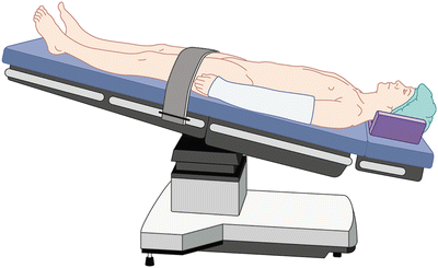

Fig. 12.2
Trendelenburg position. Note the arms tucked at the patient’s side in the neutral position with padding underneath the head and neck
Trendelenburg position is often a tilt of the bed of <20° in order to displace the viscera; however, there are many circumstances especially in urologic procedures, where steep Trendelenburg positioning of 30–45° tilt may be necessary. This steep positioning should be avoided when possible. However, if it is necessary, it is important to ensure that there is a non-sliding mattress to prevent the mattress and subsequently the patient from sliding cephalad off the OR table.
Physiologic Changes Associated with Trendelenburg Position
Cardiovascular
When placing the patient in the head-down position, there are shifts in the cardiovascular system that are both beneficial and detrimental. With a 20–45° tilt of the OR table, venous return increases, leading to an almost threefold increase in central venous pressure and an almost twofold increase in pulmonary capillary wedge pressure and pulmonary artery pressure [19]. This translocation of blood to the central compartment increases mean arterial pressure by 7–35% [20]. A reflex reduction in heart rate and peripheral vascular resistance in combination with increased stroke volume maintains a steady cardiac output [21]. The increase in venous return will also produce an increase in intracranial pressure, followed by a decrease in cerebral blood flow due to cerebral venous congestion [1].
Respiratory
The shift of abdominal viscera against the diaphragm in the supine position decreases pulmonary compliance and FRC by 20%. The decrease is further exacerbated when the patient is placed in a head-down tilt. Atelectactic areas increase, as does the mismatch between ventilation and perfusion. Hypoxemia may become an issue, especially in obese patients and those with preexisting lung disease.
Endotracheal intubation is recommended to protect against pulmonary aspiration and for utilization of positive pressure ventilation to combat atelectasis and V/Q mismatch. Elevated peak airway pressures are commonly seen once tilted head down, and various strategies should be employed to decrease these pressures during ventilation. Adjustments in inspiration to expiration ratio, respiratory rate, and tidal volume as well as utilizing pressure control ventilation rather than volume control may all be of assistance. The shift in anatomy in Trendelenburg position can also move the trachea cephalad, meaning that an endotracheal tube secured at the mouth may migrate into the right main stem bronchus, further worsening pulmonary shunt. This tracheobronchial shift may also be exacerbated during laparoscopy by a pneumoperitoneum.
These exacerbations of the Trendelenburg position on the cardiovascular and respiratory systems are transient. Returning the patient to the supine position may at times be necessary if the patient cannot tolerate the hemodynamic compromises.
Potential Complications (Table 12.2)
Brachial Plexus
The brachial plexus is subject to stretching and compression when the patient is tilted head down [22]. Kidney-shaped shoulder braces should be utilized to prevent sliding of the patient down the mattress (Fig. 12.3). With the shoulders resting firmly against these braces, there is the risk of traction on the cervical nerve roots and ensuing neuropathy [23]. In addition, when utilizing shoulder braces, abduction of the arm should be avoided. In this scenario, caudad movement of the humeral head in relation to the cervical spine may likely stretch the brachial plexus inferiorly around the humeral head [24].
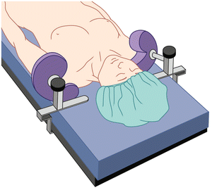
Table 12.2
Complications associated with Trendelenburg positioning
Potential complications | Recommendations |
|---|---|
Brachial plexus | Use non-sliding mattress or kidney-shaped shoulder braces |
Pad acromioclavicular joints | |
Ischemic optic neuropathy | Limit time in Trendelenburg position |
Careful management of IV fluids | |
Avoid prolonged hypotension | |
Increased intracranial pressure | Monitor head and neck for excessive edema |
Head/neck venous pooling | |
Decreased functional lung capacity and compliance | Avoid taping across chest too tightly |
Consider using alternative ventilation modes |

Fig. 12.3
Allen® shoulder restraints. Kidney-shaped padding around the acromioclavicular joints of bilateral shoulders. Avoid if arms are abducted
Ischemic Optic Neuropathy
The Trendelenburg position can lead to visual loss secondary to decreased venous return from the head. With the patient’s head below the level of the heart, increased intracranial and venous pressure can contribute to undue pressure on the optic nerve [25]. Although ischemic optic neuropathy (ION) is rarely described, it is a known complication, especially with the combination of hypotension, steep Trendelenburg, and long operative time [26]. ION is discussed in more detail in the prone positioning section later in this chapter.
Increase in Intracranial Pressure/Head and Neck Venous Pooling
Increases in intracranial and venous pressures when in Trendelenburg position for a prolonged duration can also lead to edema in areas of the head and neck. Swelling can occur of the face, eyes, larynx, and tongue and is usually more pronounced during procedures that required substantial fluid resuscitation. At the conclusion of procedures in steep Trendelenburg, it may be indicated to confirm an air leak around the endotracheal tube prior to extubation; however, this does not guarantee that an upper airway obstruction still will not occur. Continued intubation and reverse Trendelenburg following the procedure may be necessary to allow for fluid redistribution prior to extubation.
Decreased Functional Lung Capacity and Compliance
The increase in V/Q mismatch and atelectasis with Trendelenburg positioning can be compensated for in multiple ways. If patients cannot tolerate the decrease in lung capacities from the shifts during head-down positioning, it may be necessary to return them to level supine position. Interstitial pulmonary fluid may likely increase when head down; however, this change may be corrected with positive pressure ventilation when the patient is returned supine at the conclusion of the procedure. In obese patients, the additional weight from adipose tissue can dramatically effect pulmonary compliance. If possible given the surgical procedure, tilting the bed to the left or right may relieve some of the direct weight from this added tissue and improve ventilation. Lung function can be further compromised if tape has been applied too tightly across the patient’s chest and anchored to the table for added prevention against patient sliding [27].
Lithotomy Position
The lithotomy position is most frequently used for transurethral cystoscopy procedures or for open urologic procedures where access to the perineum and anus is necessary. Before repositioning the legs into stirrups, the patient’s anterior superior iliac spine should be placed over the break in the bed. The stirrups should then be anchored level with the patient’s knees and angled toward the contralateral shoulder. Once again the upper extremities can be either tucked at the patient’s side in the neutral position or abducted <90° with the arms either supinated or neutral.
When raising and lowering the legs in and out of the stirrups, it should be done in unison by at least two OR personnel so as to avoid torsion on the patient’s lumbar spine and possible dislocation of either hip. The goal of leg positioning in the stirrups is for the hips to be flexed 80–100° from the trunk and the legs abducted <30–45° from the midline (Fig. 12.4) [28]. This configuration should lead to the knees being bent until parallel with the torso. The stirrups should be padded circumferentially around the lower extremities to avoid compression injuries. When lowering the legs out of lithotomy, the knees should be brought together at midline, followed by unflexing the legs back to the supine position. Preoperative examination focused on potential limitations to hip, knee, and ankle movement should be noted.
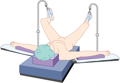

Fig. 12.4
Lithotomy position. Lower extremities are suspended in candy cane stirrups and externally rotated avoiding compression by stirrups on lateral aspect of legs
There are multiple versions of stirrups for lithotomy position including candy canes, calf rests, boots, shepherd’s crook foot straps, or Bierhoff knee crutch stirrups [2]. If using boots, the heels of the lower extremity should be flush within the boot’s footrest, and compression of the calves on the superior edge of the boot should be avoided (Fig. 12.5).
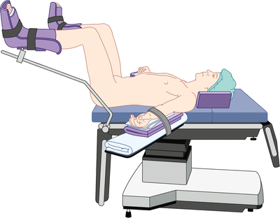

Fig. 12.5
Lithotomy position. Hips are flexed <100°; knees are flexed with legs parallel to patient’s torso. Arms are abducted <90° and positioned away from table hinge point
Exaggerated lithotomy position involves flexing the hips beyond 100°. It should be avoided due to the excessive traction it places on the sciatic and peroneal nerves. When necessary in order to perform the surgery, it should be for a limited duration of time and only during those points in the surgical procedure when it is absolutely crucial. A buttress should be placed under the lower back in these instances to relieve some of the nerve and spinal traction that the patient may experience.
Physiologic Changes Associated with Lithotomy Positioning
Cardiovascular
Elevating the legs into the lithotomy position translocates the blood volume of the lower extremities into the central compartment, increasing venous return. The acute increase in preload results in a transient increase in cardiac output leading to a potential exacerbation of congestive heart failure in at-risk patients. The autotransfusion from the lower extremities results in an increase in mean arterial pressure.
Conversely, lowering the legs at the conclusion of the lithotomy position has the opposite effect by acutely decreasing venous return and causing hypotension. The effect on blood pressure and cardiac output will depend on the volume status of the patient. Nonetheless, the effects will likely be more pronounced in patients under neuraxial or general anesthesia secondary to the intrinsic vasodilatory effects of these medications. Blood pressure should be promptly measured once the legs are lowered, in the event the patient’s fluid status cannot compensate for the translocation of blood volume back into the lower extremities.
Respiratory
Similar to the supine position, placing the legs into lithotomy position will shift the abdominal viscera cephalad into the diaphragm, decreasing lung capacities and compliance. An increase in the likelihood of pulmonary aspiration can also be seen as a result of this intra-abdominal shift. Therefore, placing an orogastric tube and suctioning the patient’s stomach contents is advised prior to converting to lithotomy.
Potential Complications (Table 12.3)
Peroneal Nerve
Neuropathy of the common peroneal nerve is the most common lower extremity neuropathy seen in the lithotomy position, accounting for 78% of lower extremity nerve injuries [29]. Potential compression of the nerve can occur when contact is made between the lateral head of the fibula and the stirrups or bar of the candy cane. It is more commonly seen in patients with low BMI, recent cigarette use, or prolonged duration >3 h [1]. The subsequent pathology seen from a peroneal neuropathy is lack of dorsiflexion of the foot, but it may also present as paresthesia or numbness. Fortunately, it is rare for this neuropathy to persist beyond 3 months, as full recovery of function and sensation is normally observed.
Table 12.3
Complications associated with lithotomy positioning
Potential complications | Recommendations |
|---|---|
Peroneal nerve | Avoid contact of fibula and stirrup |
Sciatic nerve | Avoid hyperflexion of hip and extension of the knees |
Obturator nerve | Hips flexed between 80° and 100° |
Pudendal nerve | Avoid traction and compression of legs against stirrups |
Posterior tibial nerve | Padding and appropriate angling of stirrups |
Lateral femoral cutaneous nerve | Avoid hip flexion >90° |
Saphenous nerve | Avoid compression of medial leg |
Lumbar spine torsion | Legs raised and lowered together |
Finger trauma | Arms placed on armrests away from table hinge point |
Compartment syndrome | Avoid prolonged surgical time |
Sciatic Nerve
The sciatic nerve is most susceptible to stretch injury while in the exaggerated lithotomy position. Hyperflexion of the hip in combination with extension of the knee puts the patient at greatest risk for sciatic nerve stretch neuropathy.
Obturator Nerve
The obturator nerve, which supplies motor innervation to thigh adductors, may be stretched when the patient’s hips are flexed beyond 80–100°. This amount of flexion of the thigh against the groin can injure the obturator nerve and cause it to be stretched and compressed against the pubic ramus of the pelvis as it exits the obturator foramen [30]. The obturator nerve is also at risk if the legs are first abducted and then flexed at the hip and knee when placed into the stirrups. Abducting the legs greater than 30° without concomitant hip and knee flexion places excessive stretch on the nerve.
Posterior Tibial Nerve
The posterior tibial nerve is a branch off the sciatic nerve and supplies motor and sensory innervation to the plantar surface of the foot. It is largely protected by soft tissue and muscle as it courses through the popliteal fossa, yet injury may occur if the nerve is compressed against the femoral head due to improper padding of the lithotomy boots.
Lateral Femoral Cutaneous Nerve
The lateral femoral cutaneous nerve supplies sensory innervation to the lateral thigh, and its neuropathy is typically referred to as meralgia paresthetica. Contact or compression of the lateral thigh to the candy cane stirrup rod can lead to its irritation and subsequent lateral thigh pain [31].
Saphenous Nerve
The saphenous nerve supplies sensory innervation to the medial aspect of the foot and courses along the medial side of the knee and calf. Compression from inadequate stirrup padding or by OR personnel leaning against the patient’s legs can lead to a saphenous nerve injury, most commonly from its compression against the medial tibial condyle of the knee.
Lumbar Spine Torsion
Raising and lowering the legs into the stirrups simultaneously by at least two OR personnel reduces the potential torsion placed on the lumbar spine as noted above. Patients with chronic low back pain may experience loss of their natural spine lordosis when in lithotomy, which may aggravate any underlying disease. Preoperative assessment of chronic low back pain should be noted.
Finger Trauma
With the patient’s anterior superior iliac spine placed over the crack in the bed and the arms tucked at the patient’s side, caution should be used when raising and lowering the leg support of the bed due to the proximity of the fingers to this joint. Trauma can be avoided if the arms are abducted on arm boards; however, not all procedures allow this positioning. In those circumstances, to avoid crush injury to the fingers, caution should be used when adjusting the table at its hinge points (Fig. 12.6).
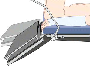

Fig. 12.6
With arms tucked, risk for finger trauma or crush injury exists when adjusting lower portion of bed in lithotomy position
Lateral Decubitus and Jackknife Position
The lateral decubitus position provides optimal surgical exposure for access to the adrenal glands, kidney, and collecting system. It is a beneficial position for removal of kidney stones located in the upper ureter and renal pelvis requiring an open procedure, as well as nephrectomies of nonmalignant disease. The patient is first anesthetized in the supine position, and then with the help of other members of the OR staff, the patient is turned to the lateral decubitus position. For extraperitoneal surgical procedures, the patient is turned a full 90°; however, if surgical exposure requires access to the intraperitoneal space, then turning 45° lateral may be adequate. Methods of maintaining the patient firmly on their side without displacement during surgical manipulation include utilizing a beanbag position immobilizer [31] or anchoring silk tape over towels placed at the patient’s shoulder and waist to the OR bed.
Jackknife is a modification of the lateral decubitus position, in which the OR table is flexed at its midpoint underneath the patient’s iliac crest. This flexing of the OR table provides stretch between the nondependent iliac crest and the costal margin on the operative side, creating a maximal surgical exposure. After flexing the table into the jackknife position, it is important to then place the table into reverse Trendelenburg until the upper torso is parallel with the ground to optimize both hemodynamic stability and tension over the incision site. If additional flexion is needed for surgery, a kidney rest can be added to the OR table apparatus [32]. The kidney rest should be anchored where the OR table breaks and placed directly under the dependent iliac crest (Fig. 12.7). This results in raising the nondependent iliac crest and promoting improved surgical exposure. Care should be taken to ensure that the kidney rest is not malpositioned underneath the flank or lower costal margin. Such malpositions can result in compression of the inferior vena cava and decreased venous return, as well as impeding ventilation of the dependent lung.
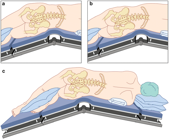

Fig. 12.7
Jackknife lateral decubitus position. Improper placement of kidney rest at (a) below the flank and (b) dependent costal margin. (c) Correct positioning below dependent iliac crest
When turning the patient to the lateral decubitus position, vigilance should be used in maintaining the head and neck in a neutral position in line with the vertebral column [33]. The head should be kept neutral by placing blankets and/or a foam doughnut beneath it for support. Failure to do so can result in lateral stretch of the neck and subsequent stretch of the brachial plexus [34]. Horner syndrome has also been reported as a possible complication of excessive lateral neck flexion due to injury of the ipsilateral stellate ganglion [35]. Attention should also be taken with the dependent eye to prevent external compression or possible corneal abrasion.
An axillary roll should always be placed and positioned beneath the patient’s rib cage just caudad to the axilla. The roll should never be located in the axilla itself, as its purpose is to distribute the weight of the thorax away from the axilla and prevent compression of its neurovascular bundle [36]. Placing the pulse oximeter on the dependent arm can be used as an indicator of axillary neurovascular compression [37]. If hypotension is recorded in the dependent arm, then axillary compression must be ruled out [38]. Periodic checks throughout the surgical procedure should also be performed to ensure that the axillary roll has not become displaced during surgery.
Stay updated, free articles. Join our Telegram channel

Full access? Get Clinical Tree


