Figure 9.1
Necrotizing Fasciitis of the anterior and lateral abdominal wall extending to the chest and axilla. There is extensive erythema and fixed staining with areas of full thickness necrosis (black eschar)
Key Features of History and Examination
Necrotizing Fasciitis (NF) is a clinical diagnosis and a life threatening condition.
History
NF tends to begin with constitutional symptoms of fever and chills. After 2–3 days, erythema is noted, and supralesional vesiculation or bullae formation ensues. Serosanguineous fluid may drain from the affected area. Necrotizing fasciitis may develop after skin biopsy; at needle puncture sites in those using illicit drugs; and after episodes of frostbite, chronic venous leg ulcers, open bone fractures, insect bites, surgical wounds, and skin abscesses. However, in many cases, no association with such factors can be made. NF may also occur in the setting of diabetes mellitus, surgery, trauma, or infectious processes.
Examination
Findings in NF may include all or some of the following clinical signs. A rapidly advancing erythema with painless ulcers appearing as the infection spreads along the fascial planes. A black necrotic eschar may be evident at the borders of the affected areas. Metastatic cutaneous plaques may occur. Septicemia is typical and leads to severe systemic toxicity and rapid death unless appropriately treated. In individuals with diabetes, crepitus is often evident, as are nonclostridial anaerobic infections. Purpura with or without bullae formation, occasionally with a lack of cutaneous erythema and heat, may be found, which does not preclude the diagnosis of NF.
Although the following features can occur with cellulitis, they are suggestive of NF:
Rapid progression
Poor therapeutic response
Blistering necrosis
Cyanosis
Extreme local tenderness
High temperature
Tachycardia
Hypotension
Altered level of consciousness
Principles of Acute Management
Laboratory Studies
Laboratory tests, along with appropriate imaging studies, may facilitate the diagnosis of necrotizing fasciitis. Although the laboratory parameters may vary in a given clinical setting, the following may be associated with necrotizing fasciitis:
WBC > 14,000/μL.
Blood urea >15 mg/mL.
Serum sodium <135 mmol/L.
Imaging Studies
Standard radiographs are of little value unless free air is depicted, as with gas-forming infections. MRI or CT scan delineation of the extent of NF may be useful in directing rapid surgical debridement
Other Tests
Excisional deep skin biopsy may be helpful in diagnosing and identifying the causative organisms. Cultures of the affected tissue obtained at initial debridement may be helpful. Gram staining of the exudate may provide a clue as to whether a type I or type II infection is present; the type influences the antibiotic therapy
Medical Therapy
This is a life threatening condition with a very high mortality rate. Management should occur within a multidisciplinary team setting. Ideally, the patient should be managed in an intensive care unit where hemodynamic parameters can be closely monitored. They will require;
Aggressive fluid resuscitation to offset acute renal failure and shock
Broad spectrum antibiotics are started, usually Clindamycin and Imipenem. This should be discussed with a microbiologist and may need to be changed when the gram stain/cultures have been reported
ITU supportive therapy with ventilation, inotropes and dialysis is also needed
Surgical Care
Once the diagnosis of NF is made, immediate surgical debridement is necessary. Surgical debridement and evaluations should be repeated almost on a daily basis until further tissue necrosis stops and the growth of fresh viable tissue is observed. If a limb or organ is involved, amputation may be necessary because of irreversible necrosis and gangrene or because of overwhelming toxicity.
These resultant wounds are best managed using a negative pressure dressing and once the tissue necrosis has stopped and the wounds are granulating they can be covered with a split skin graft.
Discussion
NF is a life threatening infection involving the superficial fascia and subcutaneous tissue. There are two types;
Type 1 involves mixed anaerobes and is usually seen in the vulnerable – young, old, immunosuppressed. It is usually an opportunistic infection
Type 2 involves Group A β-hemolytic Streptococcus (perfringens) infection which is the most common and usually affects previously fit individuals
Mortality rate is up to 50% with an often delayed presentation. There is no accepted classification system for necrotizing soft tissue infections. It is usually described on the basis of the tissue planes affected, the extent of invasion, anatomical site and causative pathogens. Deep soft tissue infections are classified either as necrotizing fasciitis or necrotizing myositis.
Key Learning Points
1.
NF is a life threatening condition with a mortality rate of 50% often with a delayed presentation.
2.
Early review by an experienced Doctor is essential.
3.
Patients should be managed within a multidisciplinary team.
4.
The mainstream of treatment involves ITU support, IV antibiotics and aggressive early surgical debridement of the affected area.
5.
Surgical debridement must be repeated on a daily basis until the NF is under control.
Clinical Case Scenario 2: Pyogenic Flexor Tenosynovitis
Case Presentation
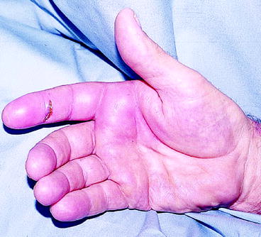
A 23 year old right hand dominant carpenter presented to the emergency department with a swollen and painful right index finger following a laceration with a Stanley knife 7 days earlier. He presented with a painful, swollen and stiff finger (Fig. 9.2) which had been gradually worsening.


Figure 9.2
Pyogenic Flexor Tenosynovitis of the right index finger showing a laceration over the middle phalanx, fusiform swelling and erythema
Key Features of History and Examination
History
Patients with Pyogenic Flexor Tenosynovitis (PFT) can present at any time following a penetrating injury. They often complain of pain, swelling, redness and stiffness in the affected finger as well as accompanying fever.
Examination
Physical examination reveals Kanaval signs of flexor tendon sheath infection, which are (1) finger held in slight flexion, (2) fusiform swelling, (3) tenderness along the flexor tendon sheath, and (4) pain with passive extension of the digit. However, Kanaval signs may be absent in some patients, such as those who have recently had antibiotics administered, early presentations and immunocompromised patients.
The differential diagnosis of flexor tenosynovitis includes the following:
Inflammatory (nonsuppurative) flexor tenosynovitis
Herpetic whitlow
Pyarthrosis
Gout
Dactylitis
Phalanx fracture
Arthritis
Principles of Acute Management
If PFT is suspected, it is important to keep the patient nil by mouth and urgently refer them to a Plastic Surgery Unit. The indication for surgical drainage includes history and physical examination consistent with acute or chronic flexor tenosynovitis. In certain circumstances when acute flexor tenosynovitis presents within the first 24 h of onset, medical management may initially be trialed. Prompt improvement of symptoms and physical findings must follow within the ensuing 12 h; otherwise, surgical intervention is necessary.
Laboratory Studies
1.
If infection is suggested, culture of the suppurative synovial fluid is mandatory prior to commencing definitive antimicrobial treatment
These cultures should include aerobic, anaerobic, fungal and acid-fast bacilli
2.
Full Blood Count
WBC count may be elevated in the presence of proximal infection or systemic involvement. WBC count is not elevated in nonsuppurative conditions
WBC count is often not elevated in immunocompromised patients.
3.
Erythrocyte sedimentation rate (ESR)
Although nonspecific, the ESR is typically elevated in acute or chronic infections and may serve as a marker to follow resolution of an infection
ESR may be elevated in cases of inflammatory FT as well
4.
Rheumatoid Factor is useful if rheumatoid arthritis is a consideration
Imaging Studies
Obtain standard anteroposterior and lateral radiographs to rule out bony involvement or retained foreign body.
Medical Treatment
If a patient presents very early medical treatment may initially be used. This includes;
1.
Broad spectrum intravenous antibiotics
2.
Elevation
3.
Physiotherapy – once PFT is under control
Surgical Treatment
Indications;
1.
No response to medical treatment within 12–24 h
2.
Late presentation
3.
Immunocompromised or diabetic patients
Surgical Procedure
Closed tendon sheath irrigation is carried out. A proximal incision is made over the A1 pulley. In the digit, either a standard Brunner incision or a midaxial incision may be utilized. The distal incision is made over the region of the A5 pulley. An appropriate size feeding tube is inserted into the tendon sheath through the proximal incision. The sheath is copiously irrigated with a minimum of 500 mL of normal saline. The wounds are left open, a splint is applied and the hand is elevated, and empiric antibiotic coverage is started while awaiting culture results.
After 24–48 h the wounds are inspected. For persisting infection, repeat operative debridement may be required. Otherwise: the wounds should be left open to heal by secondary intention and physiotherapy should be commenced. The switch from IV to oral antibiotics should be based not only on the culture results but also on the clinical examination and patient’s progress.
Discussion
PFT results from an infectious agent multiplying in the closed space of the flexor tendon sheath and culture-rich synovial fluid medium. Natural immune response mechanisms cause swelling and migration of inflammatory cells and mediators. The septic process and inflammatory reaction within the tendon sheath quickly interfere with the gliding mechanism, leading to adhesions and scarring. This can ultimately result in tendon necrosis, disruption of the tendon sheath, and digital contracture.
The most common organisms responsible for disease include Staphylococcus aureus and β–hemolytic Streptococcus. If the initial injury was caused by an animal bite Pasteurella multocida should be suspected and if a human bite Eikenella corrodens or Anaerobes.
Key Learning Points
1.
Clinical examination is the hallmark of diagnosis.
2.
Kanaval signs of flexor tendon sheath infection are (1) finger held in slight flexion, (2) fusiform swelling, (3) tenderness along the flexor tendon sheath, and (4) pain with passive extension of the digit.
3.


Kanavel’s four cardinal signs may not all be present during the early stages of the disease, in the immunocompromised and in diabetics.
Stay updated, free articles. Join our Telegram channel

Full access? Get Clinical Tree


