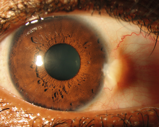
Eric D. Hansen, James T. Banta Pinguecula and pterygium are two of the most common ocular surface abnormalities with which patients visit primary care providers. Pingueculae are benign, elevated lesions of the bulbar conjunctiva, often located adjacent to the cornea. Pterygia are similarly benign and originate in the conjunctiva but have grown onto the surface of the cornea. The word pterygium derives its origin from the Greek word pterygion meaning “small wing”—an apt name because of the winglike shape the growth displays on the cornea. Both pingueculae and pterygia usually grow slowly and commonly manifest in patients 20 to 50 years of age. They are more common in lower latitudes with greater sun exposure. Pingueculae and pterygia have both degenerative and proliferative components. Pathologic analysis reveals fibrovascular proliferation and disruption of the basement membrane of the conjunctival epithelium along with basophilic and elastotic degeneration of the substantia propria. Corneal involvement in pterygia includes infiltration of the Bowman layer, a strong layer of collagen directly beneath the corneal epithelium. Disruption of this layer can lead to scarring and focal opacification of cornea. Although the pathogenesis of these entities is multifactorial and incompletely understood, exposure to ultraviolet (UV) light, particularly UVB, has been associated with development of pterygia.1,2 Heredity and chronic conjunctival inflammation also may contribute.3,4 Pingueculae and pterygia have no common systemic associations, although they may occur more frequently in conditions that confer a proclivity for skin neoplasms, such as xeroderma pigmentosum. However, despite this association and the link to UV light exposure, pterygia and pingueculae carry no significant malignant potential. In addition, because they are degenerative processes, development before the age of 20 years is very unusual. Both pingueculae and pterygia arise and grow slowly over the course of years. Common complaints on the part of the patient include dryness, irritation, redness, foreign body sensation, and itching, as well as the cosmetic appearance of the lesion itself. Severe dryness from tear film irregularity induced by the lesion may cause intermittent blurry vision. In more advanced stages of disease, pterygia may cause decreased vision by two other mechanisms: astigmatism, which is irregularity of the corneal shape as a result of the physical presence of the pterygium; and obstruction of the visual axis by extension of the lesion into the central cornea. The diagnosis of pinguecula and pterygium is often made clinically. Most lesions are detectable with the naked eye. The overwhelming majority of lesions appear in the horizontal meridian at either the 3-o’clock or 9-o’clock position, more frequently nasally than temporally. A single eye may have a pinguecula or a pterygium at both positions or a combination of a pinguecula at one and a pterygium at the other. Pingueculae appear as elevated, yellowish-white lesions immediately adjacent to but not encroaching on the cornea. They do not typically have abnormal vasculature or large feeder vessels and do not bleed spontaneously. Pingueculitis, or inflammation of a pinguecula, may occur, in which case the lesion may be pinkish with dilated blood vessels over and surrounding it (Fig. 78-1). Patients typically report increased redness and irritation in the area.
Pinguecula and Pterygium
Definition and Epidemiology
Pathophysiology
Clinical Presentation
Physical Examination
Pinguecula and Pterygium
Chapter 78