Perioperative Approach to the High-Risk Surgical Patient

Chapter 86


Perioperative Approach to the High-Risk Surgical Patient image


The changes in cellular and biochemical functions that accompany and follow major surgery differ from most others encountered in medical practice. Fortunately, the response to injury follows a predictable patterned response trajectory. These differences in normal homeostasis and the “stress” response become even more important clinically when the patient undergoing surgery is “high risk.” High-risk patients are those with preexisting conditions, such as comorbidities or advanced age, that markedly alter their physiologic reserves.


Stress Response to Acute Injury


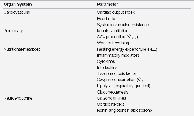
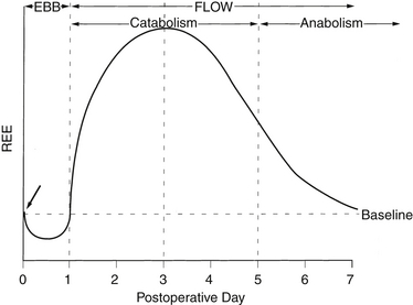
Acute injury results in a characteristic biphasic set of physiologic and metabolic changes, collectively described as the stress (or inflammatory) response (Figure 86.1). The initial period, which Cuthbertson termed the ebb phase in his original description, is associated with decreased cardiac output, a reduction in blood flow to organs other than the brain and central nervous system (CNS), hypothermia, and an overall decrease in the resting energy expenditure (REE). In modern parlance this is a stage of shock. As is most often the case with shock, the transition from the ebb phase is rapid when adequate resuscitation is provided. The subsequent period, or flow phase, is characterized by fever, an increase in blood flow to most tissues, and an increased REE. Virtually all variables associated with acute inflammation peak on or about postinjury day 2 and then decline to baseline by postinjury day 6 to 7 (Table 86.1). Deviations from this expected pattern indicate the influence of preexisting medical conditions or postoperative complications.


Hypermetabolic Phase


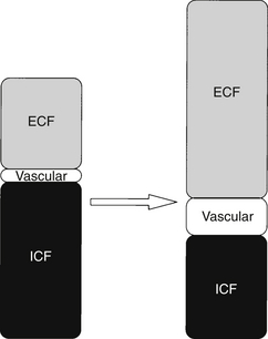
The increase in energy expenditure (hypermetabolism) that characterizes the flow phase reflects the initiation of processes to repair damaged tissue. The increase is driven primarily by the metabolic activities of inflammatory cells. These cells are obligate glucose users. A continuous supply of glucose is provided by enhanced hepatic gluconeogenesis and glycogenolysis. Skeletal (somatic) and visceral (smooth) muscles are catabolized into amino acids to be used as substrate for the synthesis of structural proteins and enzymes. The energy needs of other organs are primarily met by the oxidation of fatty acids. This global increase in metabolism is reflected in a rise in REE, oxygen consumption, and carbon dioxide production. In the flow phase, peripheral vascular tone decreases and cardiac output increases to provide flow to injured tissue and deliver nutrients to muscle and the liver. Minute ventilation rises in proportion to the increase in carbon dioxide production, keeping Paco2 in the normal range. However, vascular dilation and capillary recruitment are insufficient to support the delivery of substrate to damaged tissue. Most injured tissue is essentially avascular, and substrate delivery is primarily a function of diffusion across a concentration gradient into the interstitium. To increase substrate delivery to a wound, capillaries become “leaky” and fluid is lost to the extravascular compartment. This results in intravascular fluid depletion. To compensate there is an increase in renal salt and water retention and fluid translocation from the intracellular compartment (Figure 86.2).


Unchecked, the hypermetabolic process would eventually result in death. By the fourth to fifth postoperative day, however, likely as a direct result of neovascularization, there is restoration of blood flow to the wound and enhanced delivery of substrate that reverse the hypermetabolic process. This subdivision of the hypermetabolic phase was first recognized by Francis Moore. He called the pre-neovascularization period the catabolic phase (reflecting the catabolism of endogenous protein and loss of cellular water) and the post-neovascularization period the anabolic phase.


The anabolic phase is characterized by restitution of body cell mass; movement of water, potassium, magnesium, and phosphate back into cells; vasoconstriction; and mobilization of interstitial fluid. Clinically, there is a brisk diuresis, resolution of anasarca as the capillary leak resolves, and a decrease in serum levels of potassium, magnesium, and phosphate (resulting in the need for replacement therapy). The time required to restore the tissue depletion that results from catabolic changes is proportional to the degree of injury and typically takes weeks to months after major surgery or trauma.


The hypermetabolic flow phase is driven by the energy required for tissue repair. To date, attempts to prevent this phase have been ineffective. For example, the use of epidural anesthesia to block the sympathetic nervous system delays the onset of catabolism, but once the anesthetic is removed, hypermetabolism begins. Similarly, drugs such as beta-adrenergic blockers (which limit a patient’s ability to achieve the peak metabolic and physiologic changes of the hyperdynamic phase) can reduce the peak hypermetabolic response but subsequently prolong its duration.


Postoperative Issues


Concurrent diseases may have their greatest impact by limiting responses during the hyperdynamic phase. The magnitude of these changes depends on the extent of the initial injury as well as on preexisting diseases (Table 86.2). Three important factors have an impact on the patient’s ultimate outcome. The first factor is the ability of the patient to mount a hypermetabolic response. In some cases, this requires high levels of hemodynamic support. In others, however, the intensive care unit (ICU) physician’s role in management is simply to support the hypermetabolic response, particularly by replacing ongoing fluid losses (especially those lost into the “third space”). This allows the cardiovascular system to become hyperdynamic, which, in turn, supports blood flow and oxygen delivery to the wound and major organ systems such as the liver and kidneys.


The second factor is the ability of the ICU clinician to recognize when the patient’s response deviates from the expected pattern. The patient who remains hypermetabolic—that is, manifesting neither a brisk diuresis nor requiring replacement of potassium, magnesium, or phosphate by postoperative day 6 is not following the expected response trajectory. He or she has an ongoing hypermetabolic process that reflects a complication, most often infectious. The third factor is the extent to which preexisting disease and therapy for those disorders alter the ability of the patient to tolerate hypermetabolism or to resolve the hypermetabolic state. For example, beta-blockade may alter the patient’s ability to become hypermetabolic, thus limiting or prolonging the expected response.


Preoperative Issues: Preparing the High-Risk Patient for Surgery


Response to surgical trauma should be viewed within the context of the stress response. In this paradigm, the ebb phase is initiated by the induction of anesthesia. This leads to venous pooling, a decrease in cardiac output secondary to a loss of preload and a decrease in blood flow to all systems other than the CNS. This may be exacerbated by intraoperative blood loss, insensible fluid losses, and under-resuscitation. If fluid resuscitation is appropriate, the transition to hypermetabolism will begin intraoperatively. This imposes an additional need for fluid to accommodate the increased perfusion and capillary leak that are characteristic of the flow (hypermetabolic) phase. Preparing the high-risk patient for surgery requires anticipating this pattern and appreciating how the patient’s physiologic limitations may interfere with the fluid requirements imposed by hypermetabolism. image


The hallmark of this perioperative approach to the high-risk surgical patient is to ensure the optimal functioning of physiologic systems when they are activated in response to surgical stress. Many surgical interventions can be anticipated well in advance, allowing time to plan a course of action. When dealing with a critically ill patient, however, only hours rather than days may be available for this preparation.


Fortunately, even with limited time, a great deal can be done to support the cardiopulmonary and metabolic reserves of the patient preoperatively. Key interventions are (1) restoration of fluid volume stores, (2) correction of important electrolyte abnormalities, (3) limitation of exogenous catecholamine administration, (4) maintenance of normothermia, (5) correction of reversible impairment of pulmonary mechanics, particularly bronchospasm, and (6) initiation of other exogenous therapy, such as mechanical ventilation and metabolic support, when indicated.


Specific Perioperative Interventions


Correcting Volume Deficits


The preoperative management of the critically ill or high-risk surgical patient begins with ensuring adequate intravascular, interstitial, and intracellular fluid volumes. Because the delivery of substrate for repair of damaged tissue is accomplished in part by the development of capillary leak, fluid shifts from the intravascular and intracellular compartments into the interstitial compartment occur even in undamaged tissue. As a result, postoperative physiology will be exaggerated in the face of any preexisting deficit in intravascular or intracellular fluid volume. Further, adequate intravascular volume is essential for the development of the hyperdynamic cardiovascular response. Repleting intravascular volume permits the development of an appropriate hemodynamic response without excessive tachycardia and its associated myocardial oxygen demand.


Assessing and achieving proper fluid balance in critically ill patients can be difficult (Chapter 7). This problem is amplified in patients with major preexisting medical disorders (see Table 86.2). Additionally, some patients appear to be in fluid balance or even overloaded when, in fact, a relative intravascular volume deficit exists. For example, the use of diuretics in patients with congestive heart failure or hypertension can lead to total body deficits of water, sodium, and potassium. These may result in an inability to mount an adequate postoperative hyperdynamic response.


A number of surgical factors can be expected to exaggerate relative preoperative hypovolemia. Critically ill patients taken to the operating room from the ICU or the emergency department may have unrecognized intravascular volume deficits resulting from infection, fever, or bleeding. Patients receiving bowel preparation agents prior to surgery and those who have been nil per os (NPO) may have significant volume deficits upon presentation. The acute venodilatory and anti-inotropic effects of anesthetics can compound the hemodynamic consequences of preoperative hypovolemia, placing the patient at even greater risk. Major drops in blood pressure associated with the induction of anesthesia are often a sign of intravascular fluid depletion.


In short, the safe conduct of surgical intervention and the requirements of postoperative physiology demand that hypovolemia be avoided or rapidly corrected in the preoperative and intraoperative periods.

< div class='tao-gold-member'>

Stay updated, free articles. Join our Telegram channel

Jul 7, 2016 | Posted by in CRITICAL CARE | Comments Off on Perioperative Approach to the High-Risk Surgical Patient

Full access? Get Clinical Tree

Get Clinical Tree app for offline access