Necrotizing Fasciitis and Related Soft Tissue Infections

Chapter 66


Necrotizing Fasciitis and Related Soft Tissue Infections


In 1871, Confederate Army surgeon Joseph Jones reviewed more than 2600 cases of “hospital gangrene”—the first published description of necrotizing soft tissue infection (NSTI)—revealing a mortality of 46%. Although better understanding of the pathophysiology of these infections, directed therapy with modern antibiotics, and greater awareness of the disease among clinicians have improved the prognosis of these deep tissue infections, NSTI remains a diagnostic and therapeutic challenge with substantial morbidity and mortality.


Several factors have been implicated in contributing to poor outcomes. Perhaps the most important prognostic indicator is the time to diagnosis. Subtle and relatively benign changes of the skin overlying NSTIs commonly belie the magnitude of destruction underneath. The insidious nature of many NSTIs requires great awareness and a high index of suspicion. Potentiating the delay in the diagnosis of NSTI is its rarity. In the United States, NSTI has an incidence of ∼1000 cases per year. Thus, most ICU practitioners see only a few cases in their careers—making recognition of NSTI that much more challenging.


Soft Tissue Infections


Layers of Soft Tissue


The “soft tissue” is composed of four distinct layers of tissue: the skin, subcutaneous tissue or “superficial fascia,” deep fascia, and muscle. Dividing the soft tissue into these four anatomic layers can be helpful both descriptively and therapeutically. Infectious processes that affect the two most superficial layers are usually self-limited and can be treated effectively with nonsurgical therapy—that is, local hygiene and antibiotics. Infections of the deep fascia and muscle, which can spread rapidly and produce large areas of tissue destruction culminating in multiorgan dysfunction, virtually always require surgical therapy.


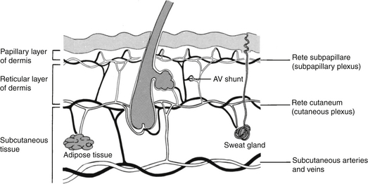
The skin is a two-layer membrane consisting of the epidermis and dermis. These layers are tightly fused above the subcutaneous tissue. When intact, the skin presents an almost impenetrable barrier to microorganisms. However, skin damaged by trauma or disease does not provide nearly as effective a defense. The blood supply of the skin runs horizontally at the junction of the dermis and subcutaneous tissue (Figure 66.1), and it is crucial for maintaining the skin’s integrity.


The subcutaneous tissue or superficial fascia, located between the dermis and deep fascia,consists of fat and loose connective tissue. Most soft tissue infections occur at this level and are commonly termed cellulitis or adipositis. The subcutaneous tissue is only loosely fixed to the deep fascia. This junction between superficial fascia and deep fascia is a potential space, called the fascial cleft. Infection can spread rapidly in this plane, impeded only where the superficial fascia is adherent to bone.


The deep fascia is a layer of strong connective tissue that overlies and separates major muscle groups. Where present, the deep fascia effectively deters the spread of infection from the superficial fascia into the muscle. Infection that extends to this layer and spreads within the fascial cleft is often termed fasciitis. Where the deep fascia is absent, for example, in the face and scalp, superficial infections can quickly spread into deeper tissues.


Skeletal muscle is the deepest layer of soft tissue and is made up of long multinucleated cells enclosed in the sarcolemma. Multiple cells are held together by a fibrous epimysium in which nerves and blood vessels run. The blood supply is extensive, with each muscle fiber receiving blood from several capillary beds. The richness of this blood supply is, in part, responsible for muscle’s high resistance to infection. When infection occurs at this level, it may be called myositis or myonecrosis.


Pathogenesis


Although soft tissue infections can originate from defects in systemic defenses, they more commonly develop as a result of local damage to the corneal layer of the epidermis, allowing microorganisms to invade. Alternatively, NSTIs sometimes originate from underneath the skin by way of a perforated viscus—notably the colon, rectum, or anus. Epidermal damage leading to NSTI can be caused by trauma as subtle as that associated with tape removal, hair plucking, or occlusive dressings that retain water and macerate the skin.


Like the epidermis, normal soft tissue is resistant to infection. Other factors influencing the development of NSTI include the size of the bacterial inoculum, local host defenses, tissue perfusion, and adjacent tissue trauma.


Another factor leading to the severity of some NSTIs is the contribution of exotoxins from some of the causative organisms. Clostridial infections are complicated by α-toxin, which causes tissue destruction and hemodynamic instability. Infections by Staphylococcus aureus and streptococcal species are complicated by a plethora of toxins. M-surface proteins enhance the bacteria’s tissue adherence and ability to evade host defenses. Exotoxins A and B cause capillary leakage and impaired blood flow. Streptolysin O and superantigen lead to a hyperinflammatory response resulting in severe sepsis or septic shock, which is often unrecoverable.


Thrombosis of deep blood vessels of the affected area is a hallmark of NSTI. However, the skin is able to receive its blood flow largely from the skin around it via the horizontal vascular plexus in the deep dermis (see Figure 66.1), rather than from the tissue beneath, leading to the NSTI’s often underwhelming external appearance.


Microbiology


Necrotizing soft tissue infections may by caused by a variety of organisms, and in many combinations. Historically, clostridia are often cited as the causative organism of necrotizing fasciitis (for example, in “gas gangrene” described in trench warfare). Although clostridial soft tissue infections still exist today, they are uncommon. Two microbial subtypes of NSTI are usually described, with the addition by some authors of a third.


Type I infections are polymicrobial and are the most common form; they are commonly found on the trunk and perineum. An average of four different organisms may be cultured from such wounds and usually include a combination of gram-positive cocci, gram-negative rods, and anaerobes. Both bacteroides and clostridia may be present. Clostridium septicum is a rare finding, but when present it is highly suggestive of a perforated colon cancer. Type I infections tend to occur in immunocompromised patients.


Type II infections are caused by a single organism, usually Streptococcus pyogenes (i.e., group A streptococcus), occasionally in association with Staphylococcus aureus, and are commonly found on the extremities, though systemic toxic shock syndrome can result. In addition, community-acquired methicillin-resistant Staphylococcus aureus (MRSA) has become more common, especially in intravenous (IV) drug users; MRSA may now be isolated in 40% of necrotic wounds. These infections tend to occur in otherwise healthy, young patients.


A type III infection has been described and includes the small subset of NSTIs caused by Vibrio vulnificus. This bacterium is encountered when open wounds are exposed to warm seawater, though necrotizing soft tissue infection by this mechanism is unusual. Type III NSTI is remarkable for its rapid progression to multisystem organ failure and death within 24 hours.


Clinical Manifestations and Differential Diagnosis


Skin Infections


Most infections confined to the skin are caused by streptococcal and staphylococcal infections. Examples of these types of infections, nonspecifically called pyodermias, include impetigo, ecthyma, and erysipelas (Table 66.1).


< div class='tao-gold-member'>

Stay updated, free articles. Join our Telegram channel

Jul 7, 2016 | Posted by in CRITICAL CARE | Comments Off on Necrotizing Fasciitis and Related Soft Tissue Infections

Full access? Get Clinical Tree

Get Clinical Tree app for offline access