(1)
Division of Pulmonary and Critical Care Medicine, Eastern Virginia Medical School, Norfolk, VA, USA
Keywords
Type 1 respiratory failureType II respiratory failureHypoxiaMechanical ventilationTidal volume ARDSnetPositive end-expiratory pressure (PEEP)Pressure controlled ventilation (PCV)Airway pressure release ventilation (APRV)Esophageal tonometryNon-invasive ventilation (NIV)Continuous positive airway pressure (CPAP)Pressure support ventilation (PSV)Respiratory rateMode ventilationAssist controlled ventilation (AC)Acute respiratory distress syndrome (ARDS)Fraction of inspired oxygen (FiO2)Pressure-volume curveAuto-PEEPTracheostomyPlateau airway pressureBarotraumaThe requirement for endotracheal intubation and mechanical ventilation is the most common indication for admission to the ICU. The most common reason for mechanical ventilation is hypoxic respiratory failure (type 1 respiratory failure). In addition, patients with hypercarbic respiratory failure (type II respiratory failure) who have failed non-invasive modes of ventilation (NIV) require mechanical ventilation. With the development, refinement and popularization NIV as a primary mode of ventilatory support (CPAP and Bi-PAP) many patients who previously would have required intubation and mechanical ventilation are now treated with NIV [1–3]. The most common indications for NIV are an acute exacerbation of chronic obstructive lung disease (COPD) and acute cardiogenic pulmonary edema (see Chap. 20). NIV is generally inappropriate for patients with severe respiratory failure due to pneumonia, aspiration pneumonitis, asthma or acute lung injury (ALI).
It should be recognized that intubation eliminates the respiratory protective reflexes (patient cannot cough effectively) and interferes with the mucociliary escalator; these effects significantly increase the risk for the development of pneumonia (ventilator associated pneumonia). In addition, mechanical ventilation is never a curative intervention, but rather provides ventilatory assistance while the underlying disorder improves. Consequently, the decision to initiate mechanical ventilation is often difficult and the clinician should weigh the risks and benefits of the intervention. It is also important to recognize that positive pressure ventilation is potentially lethal in patients with severe pulmonary hypertension (may cause severe RV failure). The decision to intubate and initiate mechanical ventilation is essentially one of clinical judgment and should be based on a number of factors including the respiratory rate, heart rate, blood pressure, signs of respiratory distress (nasal flaring, use of accessory muscles, use of abdominal muscles, grunting, etc), arterial blood gas analysis (ABG-PaO2, PaCO2, pH) and/or pulse oximetry as well as the patients co-morbidities, acute medical problem and the likely response to medical interventions.
The most common indications for intubation and mechanical ventilation are listed below:
Hypoxic respiratory failure
◦ Deliver a high FiO2
◦ Reduce shunt
◦ Apply PEEP
Hypercapnic respiratory acidosis
◦ Reduce the work of breathing (WOB) and thus prevents respiratory muscle fatigue
◦ Maintain adequate alveolar ventilation
Unprotected and unstable airways (e.g., coma)
◦ Secure the airway
◦ Reduce the risk of aspiration
◦ Maintain adequate alveolar ventilation
Other
◦ To facilitate procedure (bronchoscopy), bronchial suctioning
Think of Respiration as Two Separate Processes
Oxygenation (assessed by PaO2 and percent saturation)
Ventilation (alveolar ventilation is indirectly proportional to PCO2)
A landmark study published by the ARDSNet group (NIH ARDS Network) in 2000 demonstrated that volume-assisted ventilation (AC) with a low tidal volume (6 mL/kg of predicted body weight) was associated with a significant reduction in 28 day all-cause mortality as compared to AC ventilation with traditional tidal volumes (12 mL/kg of PBW) in patients with ARDS [4]. Such an approach is now considered the standard of care and applies to all mechanically ventilated patients, not just those with ARDS [5–7].
The predicted body weight (PBW) should be calculated on all patients undergoing mechanical ventilation
Men: PBW = 50.0 + 0.91 (height in centimeters—152.4)
Women: PBW = 45.5 + 0.91 (height in centimeters—152.4)
Regardless of the mode of mechanical ventilation the tidal volume (Vt) of all patients undergoing mechanical ventilation should target 6 mL/kg PBW and should never exceed 8 mL/kg PBW [4]. Alveolar overdistention has clearly been shown to damage normal as well as injured lungs. It is therefore important that a low Vt (known as a lung protective strategy) be used in all patients undergoing mechanical ventilation. Furthermore, it is important to use the predicted body weight (PBW) and not actual body weight for these calculations. The concept underlying this approach is that it normalizes the Vt to lung size, since lung size has been shown to depend most strongly on height and sex. For example, a person who ideally weighs 70 kg and who then gains 35 kg has essentially the same lung size as he or she did when at a weight of 70 kg and should not receive ventilation with a higher Vt because of the weight gain.
Alveolar Overdistension Damages Normal Lungs
In an observational cohort study, Gajic et al. reported that of patients ventilated for 2 days or longer who did not have ALI/ARDS at the onset of mechanical ventilation, 25 % developed ALI/ARDS within 5 days of mechanical ventilation [8]. In a multivariate analysis, the major risk factors associated with the development of lung injury were the use of a large Vt and transfusion of blood products. Interestingly, female patients were ventilated with larger Vt (per predicted body weight) and tended to develop lung injury more often. Women are generally shorter than men … this may have accounted for this finding. Similarly, in a large prospective observational study, a large Vt and high peak airway pressure were independently associated with development of ARDS in patients who did not have ARDS at the onset of mechanical ventilation [9].
The strongest evidence for the benefit of protective lung ventilation in patients without ALI/ARDS comes from two randomized studies in surgical patients. Intubated mechanically ventilated patients in the surgical ICU randomly assigned to mechanical ventilation with Vt of 12 mL/kg or lower Vt of 6 mL/kg [10]. The incidence of pulmonary infection tended to be lower, and duration of intubation and duration of ICU stay tended to be shorter for non-neurosurgical and non-cardiac surgical patients randomly assigned to the lower VT strategy. Futier and colleagues randomized 400 adults undergoing major abdominal surgery to an intraoperative ventilatory strategy of either 8–10 or 6–8 mL/kg [11]. The primary outcome was a composite of major pulmonary and extrapulmonary complications occurring within the first 7 days after surgery. The mean duration of ventilation was 5.7 h. In the intention-to-treat analysis, the primary outcome occurred 10.5 % assigned to lung-protective ventilation, as compared with 27.5 % assigned to non-protective ventilation (RR 0.40; CI 0.24–0.68; P = 0.001). This remarkable study demonstrates that even short periods of a non-lung protective ventilatory strategy are harmful.
Ventilator Variables and Modes of Ventilation
Current nomenclature related to mechanical ventilation are outdated and confusing. For example, the eighth edition of Mosby’s Respiratory Care Equipment lists 56 unique names for ventilator mode labels [12]. However, when analyzing the targeting schemes (the feedback control system the ventilator uses to deliver a specific ventilatory pattern) in detail, only about two dozen of these modes are “unique” and identifiable using six basic targeting schemes.
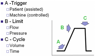
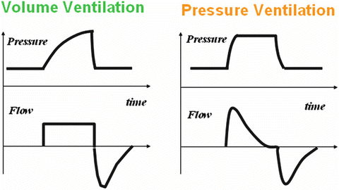
The intensivist should be familiar with a number of modes of ventilation all of which have specific indications. Standard ventilator terminology and variables are listed in Table 19.1 while initial (default) ventilator settings are listed in Table 19.2. Ventilator phase variables are illustrated in Fig. 19.1. Volume Controlled Continuous Mandatory Ventilation (VC-CMV), also termed “Assist/Control Ventilation” is the most common mode of mechanical ventilation in the ICU and can be considered a “default mode” (see Fig. 19.2). Pressure Controlled Continuous Mandatory Ventilation (PC-CMV), also termed “Pressure Control Ventilation”, is equally considered as a default/standard mode in ICUs that have a highly involved Respiratory Therapy Driven Protocol in which the exhaled VT is closely monitored (Fig. 19.2). Continuous Spontaneous Ventilation (CSV), also termed Pressure Support Ventilation (PSV) or Continuous Positive Airway Pressure (CPAP) is considered a Level 1 Evidence Based recommendations for ventilator liberation (weaning) and is commonly used in ventilator dependent patients with chronic respiratory failure (Fig. 19.3) [13]. Because the word “assist” describes the elevation of airway pressure above baseline during inspiration, PSV breath types are assisted, whereas CPAP breaths are unassisted.


Table 19.1
Ventilator terminology and parameters
Parameter | Explanation |
|---|---|
FiO2 | Fraction of inspired oxygen |
Rate | Number of breaths per minute |
Tidal volume (VT) | Volume of each breath (usually in mL) |
Sensitivity | How responsive the ventilator is to the patient’s efforts |
Peak flow | The maximum flow rate used to deliver each breath to the patient (usually in L/min) |
Inspiratory time | The time spent in the inspiratory phase of the ventilatory cycle |
I:E ratio | The inspiratory time compared to the expiratory time; I + E = total cycle time |
Flow pattern | The shape of the inspiratory flow profile representing the breath type or patient effort; it can be square wave, sinusoidal, or decelerating |
Mode | A predetermined pattern of patient–ventilator interaction. A mode can be described at various levels of detail, e.g., just specifying the control variable (volume or pressure), adding the breath sequence (e.g., VC-CMV, PC-IMV) and finally including the targeting scheme (e.g., PC-CSV with adaptive pressure targeting) |
CMV | Continuous mandatory ventilation. A breath sequence that does not allow spontaneous breaths between mandatory breaths |
CSV | Continuous spontaneous ventilation. A breath sequence consisting of only spontaneous breaths |
Cycling | The change from inspiration to expiration |
Expiration | The phase of a breath from the start of expiratory flow to the start of inspiratory flow |
Inspiration | The phase of a breath from the start of inspiratory flow to the start of expiratory flow |
IMV | Intermittent mandatory ventilation. A breath sequence that allows spontaneous breaths to occur between mandatory breaths |
Target | A predetermined goal of ventilator output such as inspiratory pressure, tidal volume, inspiratory flow or minute ventilation |
Targeting scheme | A model of the relationship between operator inputs and ventilator outputs to achieve a specific ventilatory pattern. The targeting scheme is a key component of a mode description |
Mandatory breath | A breath for which inspiration is machine triggered and/or machine cycled |
Spontaneous breath | A breath for which inspiration is both patient triggered and patient cycled |
Trigger | To start inspiration. Triggering may be machine initiated (e.g. by a preset frequency) or patient initiated (e.g., by sensing an inspiratory effort using a pressure or flow signal) |
PEEP | Positive end-expiratory pressure (usually in cm H2O) |
Table 19.2
Initial ventilator settings
Setting | |
|---|---|
Mode | VC-CMV (A/C) or PC-CMV |
VT | 6–8 mL/kg-PBW |
Rate | 12–16 min |
PEEP | 5 cm H2O |
FiO2 | 80 % |
Flow rate | 40–80 L/min |
Waveform | Decelerating |

Fig. 19.1
Ventilator phase variables

Fig. 19.2
Volume and pressure limited ventilation

Fig. 19.3
Pressure support ventilation (PSV) and CPAP
These setting should then be dynamically adjusted according to:
Plateau pressures keep less than 30 cm H2O (unless stiff chest wall disorder is present, i.e. kyphoscoliosis, morbid obesity, increased abdominal pressures, neuromuscular disease)
Arterial saturation-pulse oximetry (90–92 %)
pH and pCO2
PEEPi (intrinsic PEEP)
Flow and Pressure waveforms
Volume Controlled Intermittent Mandatory Ventilation (VC-IMV) otherwise known as, “IMV” or “SIMV” has a limited role; mainly in patients with asthma and those with unresolved respiratory alkalosis. It is the only mode of ventilation not recommended for the ventilator liberation process [13] (Fig. 19.4). Due to the ability of all ventilators to be patient triggered, it is no longer necessary to add the letter “S” to designate “synchronized” [14]. Pressure Controlled Continuous Mandatory Ventilation (PC-CMV) and Airway Pressure Release Ventilation (APRV) should be considered in patients with severe ARDS who have “failed” conventional low VT ventilation in the VC-CMV mode. In some centers APRV is considered the default mode for ALI/ARDS as well as cardiogenic pulmonary edema [15]. APRV is a useful mode in patients with atelectasis and reduced chest wall compliance.


Fig. 19.4
Synchronized intermittent mandatory ventilation (SIMV)
Ventilator Variables (See Table 19.1)
Cycling
Ventilators have traditionally been classified according to the cycling method (i.e., termination of inspiration). However, modern ventilators have microprocessors, which allow them to function in many different modes with enormous versatility. Therefore cycling is classified as machine or patient cycled. Machine cycling is defined as the inability of the patient to change the inspiratory time with inspiratory or expiratory efforts or changes to the respiratory system time constant, Therefore, examples of machine cycling include volume and time cycling.
Volume cycled. The ventilator delivers fresh gas until the preselected volume of gas is delivered. Alveolar pressure is proportional to respiratory system elastance and inversely proportional to compliance. Airway pressure is a function of volume and flow for a given elastance and resistance.
Time cycled. Inspiration continues for a preset interval, with exhalation beginning when this time interval has elapsed, regardless of airway pressure or volume delivered.
Patient cycling is the influence of the patient to changing inspiratory time by making changes in inspiratory or expiratory effort, or respiratory system time constant. Pressure and Flow cycling are examples of patient cycling.
Pressure cycled. Inspiration continues until a predetermined peak airway pressure is reached. On modern ICU ventilators this normally results in an alarm condition. The tidal volume is variable (from breath to breath) and depends on pulmonary time constant, inspiratory time, and flow rate.
Flow cycled. Inspiration continues until the inspiratory flow decays to a preset value (usually a preset flow rate or a percentage of the peak inspiratory flow rate).
Inspiratory Wave Forms
Ventilators may offer as many as three different types of inspiratory flow patterns in the VC-CMV and VC-IMV modes of ventilation. These include:
Square wave: The inspiratory flow rises to a preset level and is sustained until inspiration is cycled off.
Sinusoidal wave: The flow gradually increases and then decreases until inspiration cycles off according to the first half of a sine function. This pattern most closely mimics the normal inspiratory pattern but is not an option on most ventilators.
Stay updated, free articles. Join our Telegram channel

Full access? Get Clinical Tree


