MARINE TRAUMA
Human contact with the marine environment is becoming more frequent as recreational and commercial use of the world’s oceans increases. In addition to the hazards of drowning and cold exposure, the marine environment provides the habitat for dangerous marine fauna. Many marine animals have evolved sharp teeth and spines or venom glands for defense and predation. Encounters with marine life may result in traumatic injury or envenomation, requiring emergency medical management. Providing care for these conditions may be further complicated by the marine environment’s geographic isolation from locations with definitive health care.
Epidemiologic information typically is organized by geographic region. The most common reported U.S exposures are to jellyfish (31%), stingrays (16%), venomous fish (including lionfish, catfish, and others) (28%), and gastropods (6%).1 However, data likely favor the reporting of more severe injuries and the exclusion of common minor injuries. Human impact may be altering the geographic distribution of marine fauna as climate change affects migration patterns and artificial waterways connect previously separated bodies of water and their ecosystems.2,3
The International Shark Attack File recorded 2074 unprovoked shark attacks between 1960 and 2013, with the United States reporting the most attacks of any country and Florida reporting the most attacks of any state.4 Despite the public perception, the risk of shark attack is extremely small compared with almost any other injury. There are probably between 70 and 100 shark attacks worldwide each year, with between 5 and 15 deaths.4,5 Although the incidence of shark attacks has steadily risen since 1900, the mortality has fallen from 40% in the 30 years following World War II to current rates of approximately 10% to 20%.6 Death is usually a result of a lack of prehospital resuscitation, hemorrhagic shock, or drowning.
Other marine creatures have been reported to attack humans, typically in defense of territory rather than for feeding. The great barracuda (Sphyraena barracuda) is the only barracuda species implicated in human attacks.6 Moray eels, found in tropical to temperate waters, can inflict severe puncture wounds or lacerations, commonly to the hands of inquisitive divers. Other marine vertebrates known to cause traumatic injuries to humans include giant groupers, sea lions, seals, crocodiles, alligators, and piranhas. Some fish with sharp spines and fins (needlefish, wahoo, and triggerfish) can inadvertently injure humans. Wounds resulting from interactions with such creatures are a combination of crush injury, abrasion, puncture, and/or laceration.
Marine soft tissue infections are often polymicrobial, halophilic gram-negative infections that may be resistant to first- and second-generation penicillins and cephalosporins.7 Infecting bacteria are numerous and can vary with the environment, type of injury, and marine organism. Bacteria include staphylococci, streptococci, Aeromonas hydrophilia, Escherichia coli, Pseudomonas species, Erysipelothrix species, Chromobacterium, Edwardsiella, Shewanella, Mycobacterium species, Mycoplasma species, and Vibrio species.8,9,10 Culture results can refine antibiotic treatment. Marine-associated infections are frequently diagnosed late because the history of marine exposure or injury is poorly recalled, and patients are often initially given inappropriate antibiotics, which potentially increases the morbidity of an already virulent infection. Patients with underlying conditions such as liver disease, immunosuppression, and diabetes are more susceptible to halophilic Vibrio infections.11 Vibrio skin infections can progress rapidly from initial contact and local painful inflammation, through subepidermal bullae containing hemorrhagic fluid and vasculitis, to necrosis and small-vessel thrombosis, bacteremia, and septicemia.8,11
Encounters with sharks, skates, stingrays, barracuda, Moray eels, and giant grouper can result in major trauma. Of these animal-related injuries, shark bites are the best characterized.12,13 Shark attacks can result in neurovascular injury and significant tissue loss. Sharks typically attack the appendages (be they seals or humans), which tend to dangle lower than the head and torso while the victim is swimming on the surface. In 70% of surface swimmers, only the lower limb is involved. The upper limb can be subsequently injured when the victim tries to fend off the attacker. Sharks are unable to chew their prey and so sequentially strip it.5 In more serious attacks, substantial tissue loss and extremity amputation are common.
Stingrays possess spined tails that reflexively whip upward when stepped on or startled. These encounters typically result in foot and ankle injuries. However, serious thoracoabdominal injuries have been reported, such as the sensationalized 2006 death of naturalist Steve Irwin. Morbidity and mortality result from penetrating trauma to internal organs.14,15,16 Rarely, spine tips are retained in stingray wounds.
Victims of marine animal–associated trauma should be removed sufficiently from the water to allow immediate resuscitation. As in military settings, place a tourniquet on limbs with evidence of uncontrolled arterial hemorrhage, because the survival benefit after tourniquet placement outweighs the low risk of neurologic injury from compression.17 At the hospital, manage injuries according to institutional trauma protocols.12
However, there are two caveats for marine injuries. First, teeth and spines may be retained in bone and soft tissue and be a source of infection. Obtain plain radiographs of all injured regions to identify fractures, periosteal stripping, and retained foreign bodies. If radiographs are unremarkable but there is still a high clinical suspicion for a retained foreign body, US may be helpful. US may be particularly helpful in identifying radiolucent fragments of sea urchin spines and stingray barbs.18,19 Second, the mouths and integument of marine fauna can be heavily colonized with marine bacteria. Wounds should be swabbed and specimens sent for culture. Many marine microorganisms require special selective media for culture and sensitivity testing, so alert the microbiology laboratory that a marine-acquired organism might be present. Infections from marine microorganisms can be more serious than usual soft tissue infections. Irrigate open wounds and debride devitalized tissue. Do not suture lacerations or puncture wounds sustained in marine environments. Provide early antibiotic treatment for wounds from major trauma, in those at risk for infection, in the immunocompromised or those with hepatic disease, and for established infection (Tables 213-1 and 213-2).8,9,11,20,21 Provide postexposure tetanus prophylaxis.22 Obtain early surgical consultation for thorough debridement for suspected Vibrio vulnificus infections or any necrotizing infection. Regardless of the antibiotic choice, close clinical follow-up is essential to identify treatment failures early.
| No Antibiotic Indicated | Prophylactic/Outpatient Antibiotics | Hospital Admission for IV Antibiotics |
|---|---|---|
| Healthy patient | Late wound care | Predisposing medical conditions |
| Prompt wound care | Large lacerations or injuries | Long delays before definitive wound care |
| No foreign body | Early or local inflammation | Deep wounds, significant trauma |
| No bone or joint involvement | Wounds with retained foreign bodies | |
| Small or superficial injuries | Progressive inflamatory change Penetration of periosteum, joint space, or body cavity Major injuries associated with envenomation Systemic illness |
| For All: Staphylococci and Streptococci Coverage | Seawater Associated: Vibrio Species Coverage | Freshwater Associated: Aeromonas Species Coverage |
|---|---|---|
| First-generation cephalosporin | Fluoroquinolone | Fluoroquinolone |
| or | or | or |
| Methicillin-resistant Staphylococcus aureus coverage depending on prevalence | Third-generation cephalosporin | Trimethoprim-sulfamethoxazole |
| or | ||
| Carbapenems |
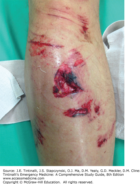
Most marine injuries and stings do not cause serious injury. Treatment depends on the agent and the seriousness of injury (Table 213-2). Coral cuts are probably the most common injuries sustained underwater and usually involve the hands, forearms, elbows, and knees (Figure 213-1). The initial reaction to a coral cut is stinging pain, erythema, and pruritus. Within minutes, the break in the skin may be surrounded by an erythematous wheal, which fades over 1 to 2 hours. With or without treatment, the local reaction of red, raised welts and local pruritus may progress to cellulitis with ulceration and tissue sloughing. The wounds heal slowly over 3 to 6 weeks.
Promptly and vigorously irrigate cuts to remove all foreign matter. Fragments that remain can become embedded and increase the risk of infection or foreign-body granuloma. Clean superficial wounds daily. Give antibiotics if infection develops (Tables 213-1 and 213-2).
MARINE ENVENOMATIONS
Venomous marine animals produce venom in specialized glands. The venom can then be applied to or injected parenterally into other organisms using a specialized venom apparatus. Venom is not a pure substance but a mixture of mainly protein and peptide toxins. The effect of a specific toxin depends on its site of action; it may be neurotoxic, hemotoxic, dermatotoxic, cytotoxic, or myotoxic. Unlike thermosTable ingestible seafood poisons, marine venoms are typically high-molecular-weight, heat-labile proteins.23
All stingrays’ tails house venomous spines.15 Inflicted lacerations allow the injection of venom. Stingray injuries cause immediate intense local pain that may radiate and last for many hours. There is often significant bleeding depending on the site of injury, and the wound may be erythematous or dusky. Systemic effects are uncommon but have been reported and relate more to the systemic response to severe pain. Submerging the affected extremity in hot water between 110°F (43.3°C) and 114°F (45.6°C) can denature the venom protein and provide pain relief within 10 to 30 minutes.24,25 Topical lidocaine can be applied for additional pain relief.
Venomous fish are found in tropical and, less commonly, temperate oceans and private aquariums. The important venomous fish include stonefish, weeverfish, scorpionfish, and lionfish. The effects of the stings range from severe with stonefish to minimal with some types of catfish and other fish with nonvenomous spines.16,26
Stonefish (Synanceia species) and scorpionfish (Scorpaenidae) occur throughout tropical and warmer temperate oceans from the central Pacific, west through the Indo-Pacific, to the East African coastline. They are a diverse group of fish, with differing habitats, swimming patterns, and ability to camouflage.16,26 The venom apparatus varies among species but most have 5 to 15 dorsal spines. Stonefish are stationary bottom dwellers usually frequenting shallow water.
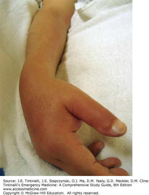
Clinical effects are characterized by immediate severe and increasing local pain, which may radiate proximally. Untreated, the pain typically peaks at 30 to 90 minutes and persists for 4 to 6 hours, but this varies considerably for different fish. The wound site usually has significant local edema and erythema (Figure 213-2). Nonspecific systemic effects such as sweating, nausea, vomiting, and even syncope may occur. Extensive tissue necrosis is not seen unless a secondary infection develops.
Weeverfish (or weaverfish) are all saltwater fish and are the most venomous fish in the temperate zone. They are found in the Mediterranean and European coastal areas and are bottom dwellers that sting when stepped on. Their five to seven envenoming dorsal spines can penetrate leather boots, producing pain severity similar to stonefish injuries. Wounds may eventually necrose.
Irrigate the wound immediately, and remove any visible pieces of the spine or integumentary sheath. Control bleeding and immerse in hot water immersion as soon as possible (Table 213-3). During the hot water soak, the wound can be explored and foreign material removed. Provide oral or parenteral analgesics as needed. The wound can also be infiltrated with lidocaine without epinephrine, or a regional nerve block can be applied to help control pain.
| Organism | Detoxification | Further Treatment |
|---|---|---|
| Penetrating Envenomations | ||
| Catfish, lionfish, scorpionfish, stingray | Hot water immersion*, topical lidocaine | Usual wound care†; irrigate with seawater or normal saline (NS) Observe for development of systemic symptoms Carefully assess for deep penetration from stingray spines |
| Stonefish, weeverfish | Hot water immersion*, topical lidocaine | Usual wound care†; irrigate with seawater or NS Stonefish antivenom for severe systemic reaction (CSL Ltd., Melbourne, Australia); be prepared to treat anaphylaxis |
| Sea snake | — | Pressure immobilization Polyvalent sea snake antivenom (CSL Ltd., Melbourne, Australia) for systemic reaction; be prepared to treat anaphylaxis Supportive care; observe for 8 h for myotoxicity and neurotoxicity; may need intensive care unit (ICU) care |
| Blue-ringed octopus | — | Pressure immobilization; flaccid paralysis and respiratory failure can develop in minutes. Provide respiratory support and ICU care |
| Cone snail | — | Pressure immobilization; observe for paralysis and respiratory failure; supportive care |
| Sea urchin and starfish | Hot water immersion*, topical lidocaine | Explore wound and remove any spines, tufts, or pincers |
| Fireworms | Topical 5% acetic acid (vinegar) | Consider topical corticosteroids Remove bristles |


