Major Vascular Procedures

Chapter 92


Major Vascular Procedures image


The care of patients with vascular disease in the intensive care unit (ICU) is challenging and requires an in-depth understanding of cardiovascular physiology and critical care. This chapter focuses on the perioperative care of the vascular surgery patient in general, as well as on particular aspects for some specific vascular procedures. Pertinent intraoperative factors will be highlighted when the condition or therapy has implications for the postoperative management.


General Approach to the Vascular Patient


Atherosclerosis is a systemic disease; therefore the majority of patients who suffer with peripheral vascular disease (PVD) also have coexisting coronary artery disease (CAD). Furthermore, patients with vascular disease frequently have a history of tobacco abuse, which often results in significant underlying respiratory disease. Hypertension, diabetes mellitus, and some degree of renal insufficiency are additional conditions commonly associated with PVD. Because these conditions are associated with morbidity and mortality after major vascular reconstruction, the preoperative evaluation and preparation of the vascular surgery patient should address both the underlying vascular disease and these associated conditions. image


As part of the preoperative evaluation, all patients require an imaging study to assess the extent of their vascular disease. Duplex ultrasound scans offer a non-invasive alternative; however, more extensive evaluation with computed tomography angiography (CTA) scans, magnetic resonance angiography (MRA), or contrast arteriography is often necessary. Whereas MRA was initially thought to avoid the risk of nephrotoxicity that accompanies iodinated contrast agents, an accumulating number of reports have documented the development of nephrogenic systemic fibrosis following the use of standard and high-dose gadolinium-containing contrast agents in patients with decreased renal function. Therefore, in addition to determining the patient’s baseline renal function, a risk benefit analysis weighing the increased diagnostic accuracy of these tests with the possibility of further renal deterioration should be performed in all patients with diminished renal function prior to the administration of intravenous contrast media.


Coronary Artery Disease


Cardiac events represent the most common cause of significant morbidity and mortality following major vascular surgery. In a large prospective cohort study involving more than 1400 patients undergoing major, nonemergent, noncardiac surgery, factors that were independently associated with myocardial infarction included preexisting CAD, age greater than 75 years, and a planned vascular surgery operation. The prevalence of CAD among vascular patients approaches 50%, and the frequency of triple-vessel CAD in these patients ranges from 15% in asymptomatic patients to 44% in those with symptoms. image


This high rate of disease leads to a perioperative myocardial infarction rate ranging from 3% to 17%. As a result, coexistent coronary disease and perioperative cardiac events account for 25% to 70% of the early and late morbidity and mortality that accompany vascular reconstructive surgery. In addition to a comprehensive history and physical exam, it is also important in the preoperative evaluation to assess the patient’s functional capacity. The inability of patients to perform their activities of daily living put them at increased risk for a perioperative cardiac event. Thus, a routine electrocardiogram should be obtained on all patients with vascular disease as part of the preoperative evaluation.


Because of the high prevalence of asymptomatic CAD, historically, most vascular surgeons prefer their patients undergo formal evaluation of cardiac function before proceeding with elective major vascular reconstruction. The most commonly used non-invasive tests to evaluate cardiac function include exercise testing, radionuclide ventriculography, echocardiography, dobutamine stress echocardiography, and dipyridamole thallium scintigraphy. Coronary angiography is performed if non-invasive testing shows evidence of myocardial dysfunction or ischemia. If angiography demonstrates significant CAD, coronary angioplasty or bypass grafting may be required before proceeding with elective vascular reconstruction. However, the relative urgency of the indicated vascular surgery must be considered because preoperative coronary revascularization has been shown to significantly postpone the intended vascular procedure.


Despite the high incidence of coronary artery disease, the benefits of routine preoperative non-invasive cardiac testing as well as pre-emptive coronary revascularization remain unclear. The positive predictive value of non-invasive imaging has uniformly been shown to be low and the test often does not provide information beyond that which can be obtained by assessing simple clinical risk factors. image


They do, however, significantly increase preoperative cost. In a large randomized trial evaluating coronary artery revascularization before elective major vascular surgery, no significant difference was noted in perioperative myocardial infarction or mortality between the groups who were randomized to either coronary revascularization or no revascularization prior to the vascular procedure. This suggests that the routine strategy of coronary artery revascularization before elective vascular surgery among patients with stable cardiac symptoms should not be universally recommended. The American College of Cardiology in collaboration with the American Heart Association published guidelines in 2007 that include a stepwise algorithm for determining preoperative cardiac risk based on patient and surgery risk factors undergoing noncardiac surgery (see http://circ.ahajournals.org/content/116/17/e418.full; accessed July 23, 2012).


Respiratory Dysfunction


Because vascular reconstructive procedures are frequently long, require a general anesthetic, and use abdominal or thoracic incisions, postoperative respiratory complications are common. Patients with vascular disease often have preexisting chronic obstructive pulmonary disease (COPD) from tobacco abuse, which increases the risk for postoperative complications as well. image


A careful preoperative history and physical examination must be performed in all patients to screen for COPD and other pulmonary diseases. All patients undergoing major vascular reconstruction should undergo chest radiography. If there is clinical or radiographic evidence of underlying pulmonary disease, arterial blood gas determinations and bedside spirometry or formal pulmonary function testing should be performed. Preoperative treatment with bronchodilators or inhaled or systemic corticosteroids is commonly used to optimize the functional status of patients with COPD before elective surgery. Finally, smokers should be strongly encouraged to abstain from tobacco use for at least 2 to 4 weeks (and preferably longer periods of time) before elective surgery. Observational studies have suggested that abstinence from smoking decreases the risk of perioperative pulmonary complications including respiratory failure, prolonged ICU admission, and pneumonia.


In patients with significant pulmonary dysfunction, it may be preferable to avoid general anesthesia, if possible. Spinal or epidural anesthesia is a frequently used alternative, particularly for lower extremity revascularization procedures. Additionally, a retroperitoneal approach can be used instead of the standard transabdominal approach for some aortic procedures. The retroperitoneal incision results in less postoperative pain and pulmonary dysfunction compared with an abdominal incision. Effective postoperative pain control is essential in order to prevent postoperative pulmonary complications. Pulmonary toilet, including coughing and incentive spirometry, is critical in the postoperative period and is made possible with effective relief of incisional pain. Epidural analgesia has become the method of choice for pain control postoperatively in patients undergoing thoracic or abdominal vascular procedures (Chapter 87).


Renal Insufficiency


Renal insufficiency is frequently present in patients with PVD. This may be the result of atherosclerotic renal artery occlusive disease, diabetic nephropathy, or uncontrolled hypertension. Vascular surgical patients with preoperative kidney dysfunction are at increased risk for developing postoperative acute renal failure (ARF). The incidence of postoperative ARF ranges from 1.7% to 25% and, when present, is associated with an increased risk of perioperative infection and mortality. image


Preventive strategies include identifying patients at risk, optimizing intravascular volume, and avoiding nephrotoxins. Common perioperative nephrotoxins include antibiotics that can be associated with acute interstitial nephritis often requiring dosage adjustments in patients with impaired creatinine clearance. Additionally, nonsteroidal anti-inflammatory drugs (NSAIDs) can cause ischemic acute tubular necrosis and acute interstitial nephritis by altering prostaglandin synthesis. Multiple studies and meta-analyses have attempted to determine if prophylactic strategies of intravenous hydration, N-acetylcysteine, or other agents reduce the incidence of contrast-induced nephropathy. Current evidence suggests that maintaining hydration with normal saline or sodium bicarbonate solutions, thereby ensuring adequate intravascular volume and sufficient renal perfusion, is the most beneficial strategy. If renal function deteriorates after a diagnostic procedure, elective surgery should be delayed to allow renal function to recover.


Monitoring


Because of the potential for hemodynamic instability, patients undergoing aortic or carotid surgery should have continuous blood pressure monitoring with an indwelling arterial catheter. The decision to place a pulmonary artery catheter should take into account the baseline cardiac status of the patient, the magnitude of the operative procedure to be performed, and the anticipated hemodynamic disturbances during and after the operation.


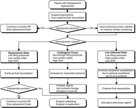
Hemoglobin, hematocrit, platelet count, and coagulation studies must also be monitored closely during the postoperative period. Blood pressure must be closely followed and tightly controlled to avoid wide fluctuations in either direction. Hemodynamic instability should be immediately assessed, and management should proceed in a stepwise and thoughtful manner image (Figure 92.E1).


Procedure-Specific Care: Abdominal Aortic Reconstruction image


Two large randomized controlled trials, the Comparison of Endovascular Aneurysm Repair with Open Repair in Patients with Abdominal Aortic Aneurysm (EVAR-1) and the Dutch Randomized Endovascular Aneurysm Management (DREAM) trial, both demonstrated endovascular aortic aneurysm repair to be a safe alternative to open repair with a clear short-term survival benefit. Although long-term results show that all-cause mortality is the same whether patients are treated with EVAR or open repair, the aneurysm-related mortality was lower in the EVAR group. However, the rate of reinterventions was significantly higher in the EVAR group. Overall, EVAR remains an attractive, less invasive option, given its shorter operative times and the avoidance of aortic cross clamping. Ongoing multicenter prospective randomized controlled trials are evaluating the role for EVAR versus open repair in healthy, asymptomatic patients.


All patients require monitoring in the ICU after open aortic reconstruction, with many patients requiring mechanical ventilation overnight. Cardiac complications are a major cause of postoperative morbidity and mortality, as previously described (see Figure 92.1). Invasive hemodynamic monitoring with a pulmonary artery catheter should be considered to direct fluid resuscitation and optimize cardiac function (Chapters 7, 8, and 11). Significant intraoperative blood loss and fluid shifts can result in hypotension in the postoperative period. Postoperative fluid requirements after aortic reconstruction may be significant because of large fluid shifts. Once the patient’s volume status has been optimized, cardiac function can be augmented with inotropic drugs if hypotension persists. Significant postoperative hypertension may result in suture line bleeding and increase myocardial oxygen consumption and therefore should also be avoided. Postoperative hypertension should be treated with short-acting vasodilators such as nitroglycerin or nicardipine for rapid titration. Occasionally, hypertension can result from postoperative pain, which, when present, should be treated accordingly (Chapter 87).


Postoperative hemorrhage is usually the result of either a technical problem or an acquired coagulopathy. Many factors contribute to postoperative coagulopathy after aortic surgery. Hypothermia, which can lead to the inactivation of clotting factors, is common with prolonged operative times and large fluid shifts. Other common causes of postoperative bleeding include decreased platelet function secondary to preoperative antiplatelet therapy, inadequate reversal of heparin with protamine sulfate, and development of a dilutional coagulopathy secondary to large intraoperative blood losses. In addition, in cases of prolonged supraceliac aortic cross clamp time, coagulation abnormalities can arise as a result of hepatic ischemia. Therefore, it is imperative to replace clotting factors and platelets as necessary and to normalize the body temperature as the first steps in the treatment of postoperative bleeding. If the patient has excessive blood loss or if the bleeding continues despite normal coagulation parameters, reoperation to look for technical causes of bleeding may be indicated.


Lower extremity ischemia after open aortic reconstruction is a well-recognized consequence of atheroemboli and occurs in 1% to 5% of patients. Limb ischemia following EVAR has also been reported to occur with a mean incidence of 5.1% and is most frequently the result of limb occlusion, although atheroembolization has also been reported. Complex aortoiliac anatomy—including tortuous, narrow, and calcified access vessels—has been proposed as a potential cause of limb occlusion following EVAR. The presence or absence of lower extremity pulses or arterial signals should be documented preoperatively and serve as a baseline to compare with in the postoperative period. The pulse exam should be reconfirmed prior to leaving the operating room and should be checked frequently in the postoperative period. The loss of peripheral pulses postoperatively demands urgent exploration and restoration of blood flow to the ischemic limb.


Clinically significant intestinal ischemia, most commonly affecting the rectosigmoid colon, occurs in 1% to 3% of patients after elective open aortic aneurysm repair. The diagnosis can be difficult because patients are often sedated and the incisional pain can mask the abdominal pain. The most common clinical signs of intestinal ischemia include bloody diarrhea, abdominal distention, peritonitis, metabolic acidosis, and hypotension. Because the sigmoid colon and rectum are almost always involved, the diagnosis can usually be made by sigmoidoscopy. A high index of suspicion is the key to early diagnosis. Cases of ischemia limited to the mucosa can be treated with supportive care, antibiotics, and close observation. Abdominal exploration, with resection of ischemic bowel and colostomy formation, is required for transmural ischemia. The overall mortality for aortic surgery complicated by clinically significant intestinal ischemia ranges from 37% to 60%. Cases of transmural ischemia and associated organ failure have been associated with even higher mortality rates. image


In emergent cases the incidence of intestinal ischemia increases to 10%. When routine postoperative colonoscopy is performed, the range of detected ischemic changes reaches 5% to 9% following elective surgery and 15% to 60% following rupture. Although EVAR has a lower rate of early postoperative mortality compared to open aortic repair, the incidence of clinically significant colonic ischemia is similar to conventional repair and ranges from 1.5% to 3%. Ischemia occurs most frequently in patients with occlusive disease of the mesenteric vessels, resulting from interruption of collateral blood flow to the intestine. The rectosigmoid colon is affected most commonly because of the frequency of disease in the inferior mesenteric and hypogastric arteries.


Acute renal failure develops in 2% to 10% of patients after open abdominal aortic aneurysm (AAA) repair and is independently associated with mortality and prolonged length of hospital stay after major vascular surgery. Hypoperfusion and atheroembolization related to aortic cross clamping as well as perioperative hemorrhage increase the risk of acute renal failure. ARF requiring hemodialysis has been reported to occur in 0.5% to 2% of patients undergoing elective AAA repair, with an associated in-hospital mortality of 25% to 66%. Regardless of repair technique, maintaining renal blood flow by optimizing fluid status and cardiac output is the most effective way to both prevent and treat postoperative renal failure. image


Endovascular aneurysm repair spares the ischemic insult to the kidneys that comes with aortic cross clamping and also is associated with less perioperative blood loss; however, nephrotoxicity can result from the intravenous contrast administered during the repair. Despite the necessary contrast load, EVAR has been associated with a 60% reduction in the risk of postprocedure ARF when compared to open aortic aneurysm repair.


Spinal cord ischemia resulting in paraplegia is an uncommon but devastating complication after infrarenal aortic reconstruction. The fundamental cause of spinal cord ischemia is a loss of adequate blood supply to the distal spinal cord. It can result from prolonged aortic clamping, intraoperative hypotension, and atheromatous embolization. Interference with the pelvic circulation such as in oversewing or exclusion of lumbar and hypogastric arteries, either by open or endovascular techniques, has also been suggested to contribute to paraplegia. image


The artery of Adamkiewicz is the major blood supply to the lower spinal cord and usually arises from a branch of the intercostal artery between T8 and Ll. Occasionally this artery may originate from the infrarenal aorta, accounting for the rare occurrence of spinal cord ischemia after infrarenal aortic reconstruction.


If a patient develops paraplegia after aortic reconstruction, magnetic resonance imaging of the spinal cord should be performed immediately to rule out an epidural hematoma as a cause of the neurologic decline. If present, immediate neurosurgical consultation is required for decompressive laminectomy. If there is no epidural hematoma, efforts should be focused on therapies to maximize blood flow to the spinal cord. Augmentation of blood pressure with vasoactive agents will increase the perfusion pressure to the spinal cord. If rapid improvement is not seen, consideration should be given to placement of a lumbar drain catheter to reduce the pressure surrounding the spinal cord.
< div class='tao-gold-member'>

Stay updated, free articles. Join our Telegram channel

Jul 7, 2016 | Posted by in CRITICAL CARE | Comments Off on Major Vascular Procedures

Full access? Get Clinical Tree

Get Clinical Tree app for offline access