Major Tissue Flaps

Chapter 91


Major Tissue Flaps image


Patients undergoing a wide variety of major flap reconstructions warrant postoperative monitoring in an intensive care unit (ICU) setting to monitor flap viability, provide adequate hemodynamic support, and rapidly recognize compromised perfusion. Because early recognition of malperfusion can result in the salvage of autografts in most patients, the ICU clinician must be familiar with the early signs and symptoms of impending ischemia.


Flap Types


A flap refers to a surgically created peninsula or island of tissue that is at least partially detached from its original site and transposed to an adjacent or distant site. It is a commonly used technique to repair defects in plastic surgery. Tissue types for flaps include skin and subcutaneous tissue, fascia, muscle, bowel, omentum, or bone, or combinations of several tissues, such as a myocutaneous flap. The anatomic vascular supply to these flaps influences the method of flap transfer and dictates which parameters are necessary to monitor postoperatively to ensure flap viability.


Skin (and its subcutaneous tissue) that is transposed to an adjacent area without being detached is a skin flap. Generally, the length of a skin flap should equal its width to provide adequate perfusion. If there is no known vessel running within the flap, it is a random flap. If there is a vascular pedicle running through the flap, either in the subcutaneous layer or just above the fascial plane, it is an axial pattern flap, and its length can be significantly longer than its width. If the skin receives its blood supply from the underlying muscle via perforating vessels, it is a myocutaneous flap, and the skin can survive as an island. image


Muscle or myocutaneous flaps, when mobilized with preservation of the vascular supply and transposed to the desired site without disrupting their vessels, are pedicled flaps. They remain attached to the patient and are limited by the available regional tissues that can be transferred to the site to be reconstructed. Their ability to reach the defect are limited by the pedicle length. Alternatively, when the muscle or myocutaneous flaps are moved to distant sites and their vessels are divided and reanastomosed to recipient vessels, they are referred to as microvascular free flaps. image


Complications of Flap Surgery


Both the recipient and the donor sites of flaps are subject to the problems of any operative wound, such as bleeding, hematoma, suture line dehiscence, infection, and localized edema (Table 91.1). Because many patients receive perioperative anticoagulation, hematoma formation is a distinct risk because flap “harvests” result in large raw surfaces at the donor site. image


Postoperative edema can severely compromise flap perfusion and stress suture lines. The operated site should be elevated above the level of the heart, if possible. The use of corticosteroids during flap harvest and early postoperatively to decrease flap edema remains controversial.


Microvascular dissection of an island of skin and fascia based on a deep vessel that spares the underlying muscle has been termed a perforator flap. Skin grafts are not considered to be flaps because they survive initially by diffusion rather than by direct perfusion.


Other tissues can also be transferred as pedicled flaps or as free flaps, including omentum, fasciocutaneous flaps (radial forearm, anterolateral thigh, and transverse scapular flaps), bone (fibula, scapula), and bowel (jejunum). The success of the operation depends on establishing a reliable arterial inflow and venous outflow.


Replantation usually involves the reattachment of a finger or thumb: scalp, ears, lips, hands, feet, penises, and rare facial avulsions have also been salvaged. If the amputation is incomplete, but the vessels are transected, the repair is a revascularization. As with a microvascular free flap, success depends on the restoration of adequate arterial and venous flow.


In the ICU, drains should be “stripped” hourly—that is, the drain is compressed manually along its length in a proximal-to-distal direction to remove clots and enhance drainage. The aim of “stripping” is to decrease the risk of hematoma and seroma formation and to promote obliteration of the donor site dead space. An unexplained drop in hemoglobin may be secondary to unrecognized donor site bleeding. Hematoma formation at the recipient site can occlude venous outflow, leading to venous congestion and flap loss.


Flap ischemia is the most devastating complication after flap surgery and can be due to arterial or venous obstruction, or both. In the case of pedicled flaps, ischemia may result from torsion of the vascular pedicle, increased tension on the pedicle as the tissues swell, or improper postoperative positioning that stretches or occludes the pedicle. The surgeon should clearly specify any restrictions in activity or positioning in the ICU postoperative orders. With microvascular free flaps and replantations, flap ischemia in the first 48 hours may be due to a technical problem that is correctable, and a prompt return to the operating room for reexploration should be considered.


Flap Monitoring: Subjective Methods


Color


A skin flap or a skin island of a myocutaneous flap (Figure 91.1) should be the same color (or slightly more pink) as the adjacent skin from which it was harvested. If it is hyperemic and purple, however, the venous outflow may be occluded. If it is mottled or extremely pale, the arterial inflow may be compromised. Of note, skin color changes in darkly pigmented patients may be difficult to discern.
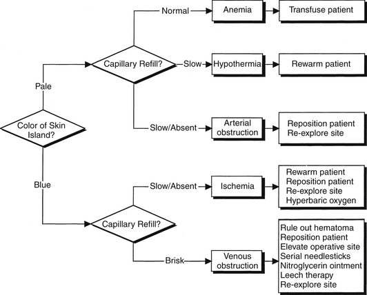


A monitoring flap is a small externally visible portion of a buried flap that serves as an indicator of the vascular status of the deeper flap. It is usually a small skin island but may also be a segment of externalized jejunum. The external component should be at least 1 × 2 cm in size to permit accurate color assessment.


In contrast to skin flaps, color is not a reliable indicator for skin-grafted muscle flaps, because the skin graft has no initial perfusion.


< div class='tao-gold-member'>

Stay updated, free articles. Join our Telegram channel

Jul 7, 2016 | Posted by in CRITICAL CARE | Comments Off on Major Tissue Flaps

Full access? Get Clinical Tree

Get Clinical Tree app for offline access