Fig. 25.1.
Trocar placement and operative positioning.
The lumbar plexus should be defined before any neurectomy is performed (Fig. 25.2) [18]. The subcostal nerve can first be identified at the T12 costal margin (Fig. 25.3). The iliohypogastric and ilioinguinal nerves, frequently sharing a common trunk, can then be seen overlying the quadratus muscle at L1 (Fig. 25.4) [19, 20]. The lateral femoral cutaneous nerve originating at L3 is identified lateral to the psoas, crossing the iliacus muscle below the iliac crest (Fig. 25.5). The femoral nerve can also be found lateral and deep to the psoas muscle, but does not require specific dissection. The dissection is then continued toward the groin where the genitofemoral nerve trunk can be noted running over the psoas muscle (Fig. 25.6). Similar to the iliohypogastric and ilioinguinal nerve trunks, the genital and femoral nerve trunks have considerable variability and often have separate trunks. If the dermatomal distribution of the femoral branch of the GFN is not affected, a separate femoral trunk should be preserved when found. Of note, the ureter and iliac vessels should be identified medial to the psoas and protected (Fig. 25.7).
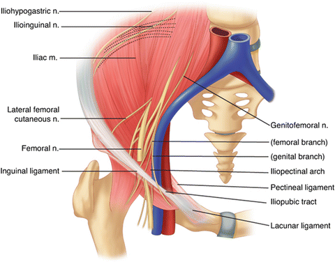
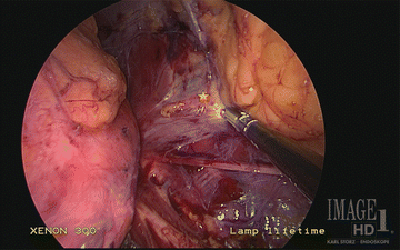
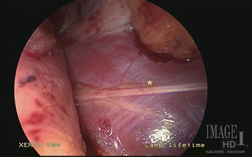
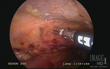
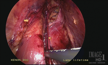
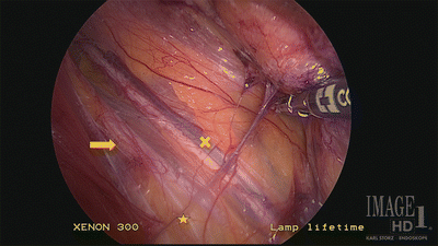
Fig. 25.2.
Retroperitoneal lumbar plexus (From Wagner et al. [18], with kind permission ©McGraw-Hill Education).
Fig. 25.3.
Subcostal nerve trunks and 12th rib at T12 level (star). Ilioinguinal/iliohypogastic nerve trunk caudal.
Fig. 25.4.
Iliohypogastric and ilioinguinal nerve trunks over quadratus lumborum muscle at L1 level (star). Subcostal nerve and 12th rib cephalad.
Fig. 25.5.
Lateral femoral cutaneous nerve trunk at L3 level (star).
Fig. 25.6.
Genitofemoral nerve trunk over psoas muscle (star). Femoral nerve lateral to psoas muscle (arrow).
Fig. 25.7.
Relationship between ureter (X), iliac artery (arrow), and genitofemoral nerve trunk over psoas muscle (star).
After all structures have been clearly delineated, the iliohypogastric and ilioinguinal nerves can be resected over the quadratus muscle. With regard to the cut nerve, our preference is to place a clip proximally and distally to close the neurilemma. This theoretically helps to avoid neuroma formation and allows for radiographic identification of the cut nerve if future proximal interventional blocks are needed. The genitofemoral nerve trunk is subsequently clipped and resected over the psoas muscle in a similar fashion. A transabdominal approach may alternatively be used to access the same anatomic planes but requires medial rotation of the viscera and more operative ports.
Outcomes
In our prospective series, 42 patients who presented with chronic inguinodynia not controlled with conservative pain management therapies underwent laparoscopic triple neurectomy (Fig. 25.8). The mean numeric pain scores were significantly reduced (baseline score 8.4) on postoperative days 1 (score, 3.4; p < 0.001), 7 (score, 2.8; p < 0.001), 30 (score, 2.4; p < 0.001), 90 (score, 2.1; p < 0.001), and 180 (score, 1.9; p < 0.001 [17]. Thirty-four patients have been followed to 12 months (pain score 1.5; p < 0.01), and 20 have been followed over 2 years (pain score 1.1; p < 0.01). Narcotic dependence was seen to decrease and the activity level of patients increased. All patients reported numbness as anticipated in the distribution of neurectomy. Fourteen (33 %) had transient hypersensitivity consistent with deafferentation, with four patients (9 %) having persistent symptoms greater than 3 months. Seven experienced residual meshoma pain with four of them undergoing a subsequent reoperation for mesh removal. Orchialgia was not improved as expected since paravasal nerves need to be resected to address this problem [16].
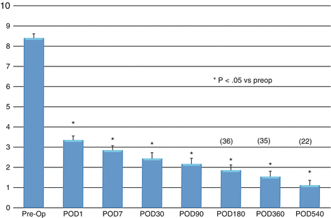
Fig. 25.8.
Laparoscopic triple neurectomy outcomes: VAS scores.
Discussion
With the advances in technique of tension-free inguinal hernia repair, chronic groin pain now surpasses recurrence as the most common long-term postoperative complication. This debilitating condition is a result of nociceptive and neuropathic factors. Given the lack of clear discrimination between the two types of pain, confounded with variables such as excitatory coupling between sympathetic and afferent nociceptive fibers, deafferentation hyperalgesia, pain centralization, and neuroplasticity, as well as patient-related factors, prevention of this complication is of key importance [9, 21].
Nociceptive pain can be minimized with gentle handling of tissues and with the use of local anesthetic to decrease the formation of nociceptive molecules. Neuropathic pain can be decreased by meticulous identification and protection of nerves to avoid injury and their direct contact with mesh, which ultimately changes the structure of their fibers. Doing so has been shown to reduce the rate of postherniorrhaphy chronic pain from 5 to 8 % to a fraction of 1 % [20]. Understanding inguinal and preperitoneal groin neuroanatomy as well as the pathophysiology of inguinal pain helps to guide good operative technique in all methods of inguinal hernia repair. Prevention and avoidance of injury at the time of the original operation are of paramount importance.
Related posts:
 Introduction to Primary and Secondary Groin Pain: What Is Inguinodynia?
Prevention of Pain: Optimizing the Laparoscopic TEP and TAPP Techniques
Prophylactic Neurectomy Versus Pragmatic Neurectomy
Patient with Referred Hip Pain
Introduction to Primary and Secondary Groin Pain: What Is Inguinodynia?
Prevention of Pain: Optimizing the Laparoscopic TEP and TAPP Techniques
Prophylactic Neurectomy Versus Pragmatic Neurectomy
Patient with Referred Hip Pain
 Chronic Pelvic Pain in Women
 Groin Pain Etiology: Hip-Referred Groin Pain
Stay updated, free articles. Join our Telegram channel

Full access? Get Clinical Tree


