Fig. 54.1
A graphical representation of the activated pathway during this test. The SCS lead stimulates the dorsal column at a level that is strong enough to activate enough fibers to excite the alpha motor neuron and generate a CMAP at the muscle. It should be noted that during normal pain therapy the levels of stimulation are much lower and thus no motor activation since not enough fibers in the specific motor pool are driven to excitement
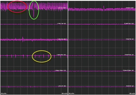
Fig. 54.2
An example of the different types of responses is noted with this technique. The response in the red circle is artifact from the stimulation device. The response in the green circle is from EKG artifact. The responses in the yellow circle are compound muscle action potentials generated by the anhidrotic activation of the alpha motor neuron pool for this muscle group
Methods
Bilateral, simultaneous free running EMG (Fig. 54.2) activity was recorded via two subdermal needles (Rhythmlink model RLSND121-2.5, Columbia, SC or Cardinal Health (Nicolet) model 019-409900, Madison, WI) placed into muscle bellies 1–2 cm apart from each other. For cervical leads , the following muscles were studied: (1) trapezius, (2) deltoid, (3) biceps brachii, (4) triceps, (5) flexor carpi ulnaris (FCU), (6) extensor carpi ulnaris (ECU), (7) abductor pollicis brevis (APB), (8) abductor digiti minimi (ADM), (9) gastrocnemius (gastroc). For the upper limb, biceps brachii and triceps are referenced, FCU and ECU are referenced, and APB and ADM are referenced. For thoracic leads , the following muscles were studied: (1) iliopsoas/adductor longus, (2) vastus medialis (Q), (3) tibialis anterior (AT), (4) gastroc (and/or soleus), (5) abductor hallucis, (6) paraspinal (rhomboid and/or erector spinae and/or trapezius—ultimate decisions on which muscles were studied may have depended on the level and amount of adipose tissue), (7) rectus abdominis (or sometimes external oblique depending on amount of adipose tissue). Needle leads were taped to the skin with either silk tape or Tegaderm and the wires secured with a piece of silk tape or a Tegaderm, 5–10 cm from the needle to act as a strain relief. All wires were run to the foot of the bed and connected to the EMG recording system amplifier. A ground pad was placed on the knee, lateral thigh, or chest. For thoracic SCS leads, only lower limbs were examined. For cervical leads, primarily only upper extremities and the trapezius were examined, but on some occasion, both upper and lower extremities were included.
Initial amplifier settings were as follows: (1) gain 100 uV/div, (2) high-cut filter (low pass) 3000 Hz, (3) low-cut filter (high pass) 30 Hz, (4) sweep 200 mSec/div. For spinal musculature, abdominal muscles, and trapezius muscles, the filter bandpass was narrowed to 100 Hz–500 Hz in order to reduce proximity stimulation artifact saturating channels. High- and low-cut filters may also require adjustment, depending upon noise levels in the operating room.
During the testing session, the gain of specific channels was adjusted during testing to account for dynamic range changes (i.e., the number of motor units activated and also the artifact amplitude). As stimulation is increased (see later), channel gains were reduced. Since the goal was to locate real EMG activity, and not the specific amplitude of this activity, such an approach was deemed to be acceptable.
Stimulation was applied through the SCS electrodes. Quadrant testing was performed (Fig. 54.3) on all lead types, except single column leads, wherein cranial (anode) and caudal (cathode) pairings could determine orientation. Stimulation was applied via the specific lead manufacturer’s screening device (Boston Scientific “Bionic Navigator”, Valencia, CA, Medtronic model 8840, Minneapolis, MN, St. Jude model 6850, Plano, TX). The lead was connected to the device via the screening cables, which were also manufacturer specific. Impedances were tested to make sure the device was working and properly connected.

Fig. 54.3
Graphical representation of the stimulation paradigms used for each lead type. The first lead on the left shows the sequence that is used with the left most electrode pair being the first tested and then sequentially going around the lead testing each electrode pair in cranial to caudal/left to right order. A similar pattern is used for the other electrodes
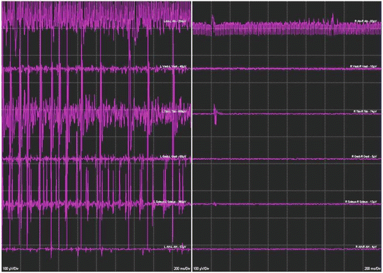
Initial stimulation testing parameters were 60 Hz and 210 μs. The amplitude was then slowly raised in 0.5 V or 0.5 mA increments until EMG activity was noted (Fig. 54.2) in any channel. Amplitude was then increased in 0.5 V (mA) increments further, until a new location of activity was detected. This process was continued until either one of three things occur: (1) the stimulator reached its maximum output; (2) both sides activated all muscle groups; or (3) one side was “completely” active, and then the stimulation was raised 1.0 V (mA) beyond this level (Fig. 54.4). “Completely” active means that all muscles on one side were firing. It is important to be able to differentiate the noise (red (stimulation artifact) and green (EKG artifact) circles in Fig. 54.2) from the actual EMG response (yellow circle in Fig. 54.2).
 Pain in the Stroke Rehabilitation Patient
Pain in the Neuromuscular Disease Rehabilitation Patient
Basic Psychopharmacology for the Treatment of Pain in the Rehabilitation Patient
Intrathecal Therapy for the Treatment of Pain in the Rehabilitation Patient
Pain in the Stroke Rehabilitation Patient
Pain in the Neuromuscular Disease Rehabilitation Patient
Basic Psychopharmacology for the Treatment of Pain in the Rehabilitation Patient
Intrathecal Therapy for the Treatment of Pain in the Rehabilitation Patient
 Intra-articular Joint and Bursa Injections for the Treatment of Pain in the Rehabilitation Patient
 Chiropractic Medicine for the Treatment of Pain in the Rehabilitation Patient


Related posts:
Pain in the Stroke Rehabilitation Patient
Pain in the Neuromuscular Disease Rehabilitation Patient
Basic Psychopharmacology for the Treatment of Pain in the Rehabilitation Patient
Intrathecal Therapy for the Treatment of Pain in the Rehabilitation Patient
Intra-articular Joint and Bursa Injections for the Treatment of Pain in the Rehabilitation Patient
Chiropractic Medicine for the Treatment of Pain in the Rehabilitation Patient
Stay updated, free articles. Join our Telegram channel
Full access? Get Clinical Tree
