Fig. 35.1
Stimulus-triggered EMG for the detection of pedicle wall breach. Electrical stimulation of hole (a) with low current activates the adjacent nerve root, evoking a CMAP response, representing a pedicle wall breach. Stimulation of the screw at hole (b) demonstrates high impedance to current through a layer of cortical bone, as no evoked CMAP is produced. This represents correct screw position. Reproduced with permission from Husain [9]
Many studies have demonstrated the value of pedicle screw EMG monitoring for the protection of nerve root and neural tissue during lumbar pedicle screw placement [10–12]. Bindal et al. [13] acquired triggered EMG recordings by stimulating the pedicle access needle and pedicle tap during TLIF. Their findings altered the trajectory of the pedicle access needle in 76.2 % of the procedures, and they felt this had led to a safer pedicle cannulation.
Raynor et al. [14] reported on the use of triggered electromyogenic stimulation (TrgEMG) in 1078 spine procedures with lumbar (L2–S1) pedicle screw placement. An ascending method of constant current stimulation was applied to each screw in order to obtain a CMAP from lower extremity myotomes. They concluded that the probability of detection of a medial wall breach by a pedicle screw increases with decreasing EMG thresholds. The probability of detecting medial wall breach with a TrgEMG stimulation threshold of greater than 8 mA was 0.31 %. A threshold of 4.0–8.0 mA yielded a probability of 17.4 %, while less than 4.0 was 54.2 %. A TrgEMG threshold of 2.8 mA had a specificity of 100 % but a sensitivity of only 8.4 %. The authors concluded that TrgEMG is a useful adjunct during screw placement, but should always be used in conjunction with some other form of monitoring.
At the same time, numerous studies have suggested that evoked CMAPs from muscles innervated by lumbar nerve roots, with stimulus thresholds of less than 4–6 mA, are suggestive of a pedicle wall breach [15–17]. Parker et al. [18] concluded that using a 5-mA stimulus threshold TrgEMG had a very high specificity but low sensitivity. Sensitivity increased with increasing stimulation at the cost of a greatly lowered specificity. It should be noted that during monitoring, false-negative results could occur when stimulating only the mobile crown of a polyaxial-type screw [14]. Anderson et al. [17] stressed the importance of stimulating the screw either at the hexagonal port or directly at the screw shank to avoid these false-negatives.
Glassman et al. [19] demonstrated with postoperative CT that lumbar pedicle screw stimulations requiring more than 15 mA to obtain a CMAP were 98 % accurate in determining that the screw was located correctly in the pedicle.
Lumbar Interbody Fusion
Lumbar interbody fusion is a commonly utilized treatment for discogenic back pain (secondary to a degenerative, herniated, or inflamed disk) and spine instability. There are several different variations of the procedure, including posterior lumbar interbody fusion (PLIF), transforaminal lumbar interbody fusion (TLIF), anterior lumbar interbody fusion (ALIF), and extreme lateral interbody fusion (ELIF).
Posterior Lumbar Interbody Fusion (PLIF)
With this surgical technique, a midline posterior incision is made, the spinal muscles are separated and retracted, and the spine laminae and a small portion of the facet joints, which lay directly over the nerve roots, are removed. The affected disks are removed and a bone graft, allograft, or biomechanical spacer implant with cage is inserted into the disk space to promote fusion of the adjacent vertebrae. The spine is then stabilized with additional instruments (rods, screws, wires). Bose et al. [20] retrospectively analyzed 61 patients having PLIFs who were monitored with continuous EMG and triggered CMAP responses from muscles innervated by nerve roots adjacent to placed pedicle screws. Twenty-one percent of patients had sustained neurotonic activity or an evoked CMAP with a current intensity of less than 7 mA during pedicle screw stimulation prompting an alert and reposition of a pedicle screw. It was their opinion that MIOM minimized postoperative neural deficits.
Transforaminal Lumbar Interbody Fusion (TLIF)
During TLIF procedures , the spine is approached from the side of the spinal canal through a paramedian posterior incision. This modified posterior approach reduces the amount of surgical muscle dissection and minimizes nerve manipulation required to gain access to the vertebrae, disk, and nerves. Laminectomy, discectomy, facet fusion, and posterolateral spine fusion and instrumentation are typically performed. Disk material is removed from the spine through the right, left, or both sides of the spinal canal, and typically, a bone graft (bone block or cage implant) is inserted. Spinal strength and stability is then achieved with screws or rods. Nerve root retraction is often needed to place the interbody device. Real-time detection of neuropraxia can be achieved with a combination of continuous EMG recordings and evoked CMAP threshold responses following electrical stimulation from pedicle screws or screw holes [13]. Bindal et al. [13] reported the use of continuous electrical stimulation of the pedicle access needle with a 7-mA current during placement of 105 lumbar pedicle screws in minimally invasive TLIF procedures in order to assess safe screw placement. Current was held constant as the needle was placed percutaneously at the lateral portion of the determined transverse process, walked medially to the junction of the transverse process and the facet joint, and then inserted into the pedicle. At this point, anteroposterior fluoroscopy was also used to confirm needle laterality. The 7-mA threshold resulted in 0 % incidence of EMG activation of the tap with a current of less than 15 mA. Detection of EMG at a current of 7 mA or less signaled close proximity of the medial nerve root resulting in lateral alteration of the needle trajectory in 76.2 % of their cases. (The author does state that there may have been false-positives. However, the 7-mA threshold was selected to yield a minimal false-negative rate.) With this method of percutaneous pedicle screw placement, they reported 0 % incidence of malpositioned hardware.
Anterior Lumbar Interbody Fusion (ALIF)
During ALIF , an incision is made in the lower abdominal area, and the abdominal muscles and vessels are retracted, allowing access to the front of the spine. When performing this procedure, particularly at the L4–L5 level, there is 5 % risk of both acute and delayed vascular insult secondary to tears and laceration of the aorta, common iliac and vena cava vessels, or retraction of the iliac arteries for adequate exposure of the disk space [21]. Venous injury occurs more frequently than arterial and is most commonly caused by retraction on the great vessels. Vascular injury is reported at a higher incidence in laparoscopic versus open ALIF [21]. Vascular tear or laceration is usually immediately recognized and repaired. However, ischemic injury due to retraction of vessels and possible thrombus occlusion may go undetected without continuous intraoperative monitoring. Undetected injury may result in a postoperative sensory or motor deficit, pain, and in some cases, mortality [22–26]. Monitoring techniques that can pick up lower extremity vascular occlusion include lower extremity vessel palpation, pulse oximetry, and spinal cord SSEP monitoring [24, 26, 27]. Nair et al. [28] report a case of iliac artery injury during an L3–S1 ALIF procedure that was detected with MIOM. On three different occasions during the procedure there were changes in tibial nerve SSEPs, abductor hallucis MEPs, or both, which corresponded with decreased pulses in the left leg following compression of the iliac artery with either a retractor or an L4–L5 spacer. Following notification to the surgeon, compression was released on all occasions, and blood flow was restored (Fig. 35.2). Similarly, Yaylani et al. [29] describe in a retrospective cohort study SSEP changes coinciding with retractor placement that subsequently resolved after surgeon notification and adjustment of the retractor during ALIF.
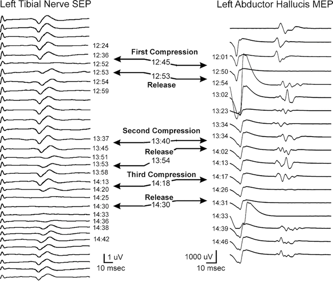
Fig. 35.2
Three compressive events during ALIF , with both SSEP and MEP signal decreases noted in the first and third compressions, and only SSEP decrease noted during the second compression. Reproduced with permission from Nair et al. [28]
Isley et al. [22] depict another case of left common iliac occlusion after anterior interbody fusion that was detected with SSEP Intraoperative monitoring. No changes in SSEPs were noted during the ALIF, and no EMG discharges suggestive of nerve root irritation were noted during the discectomies, partial corpectomies, distraction, or instrumentation. However, during closure, there was a gradual decline and loss of the subcortical and cortical SSEP waveforms. Palpation and Doppler examination verified loss of pulses in the extremity. Following vascular consult, thrombectomy and patch angioplasty of the left common and external iliac arteries was performed, perfusion was restored, and SSEP waveforms recovered. This case demonstrates the importance of continued monitoring until closure during this procedure (Fig. 35.3).
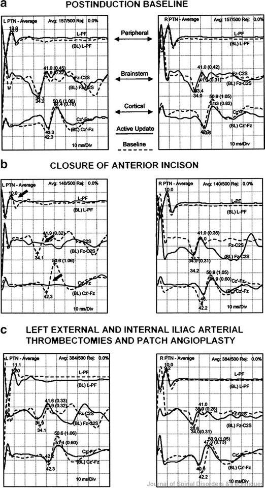
Fig. 35.3
Within each part figures are pairs of SSEP waveforms recorded at the popliteal fossa (top traces), brainstem (middle traces), and sensory cortex (bottom traces) for the left and right legs after stimulation of the PTN (labeled L PTN and R PTN, respectively) during three surgical events. Baselines are shown in (a). For the waveforms recorded to left PTN stimulation in (b), there was a loss of the dominant components at all recording sites due to thrombotic occlusion of the left common iliac arterial bifurcation. (c) Recovery of the SSEP waveforms after thrombectomies of the left common iliac arterial bifurcation. Reproduced with permission from Isley et al. [22]
Extreme Lateral Interbody Fusion (ELIF/XLIF)
The ELIF, also known as XLIF (Nuvasive, San Diego, CA) or DLIF (Medtronic, Minneapolis, MI), is a less invasive procedure with a smaller incision; however, a high incidence of lumbar plexus traction neuropraxia has been reported [23]. A flank incision is made with the patient in the lateral position, and the disk space is accessed by blunt dissection through the psoas muscle and traversing lumbar plexus. Once the disk space is entered, the disk is removed, and a new implant is inserted. During surgical dissection through the psoas muscle, traction on the lumbar plexus by the expandable cannulas used to perform the ELIF can cause a neuropraxia of the genitofemoral nerve with symptoms of thigh or groin numbness. The use of EMG monitoring using the EMG activity from muscles innervated by the lumbar nerve roots and plexus at risk during the surgery may decrease the incidence of nerve injury. The EMG-based Neurovision system (Nuvasive; San Diego, CA) incorporates a surgical dissection tool used by the surgeon that has a tiny stimulation electrode on the tip that provides the surgeon real-time continuous feedback on whether plexus structures are stimulated when dissecting through the psoas muscle.
Related posts:


Stay updated, free articles. Join our Telegram channel

Full access? Get Clinical Tree


