Fig. 42.1
Representative transcranial electric motor-evoked potentials , ulnar nerve somatosensory-evoked potentials (SSEPs) , and posterior tibial nerve SSEPs recorded during neuroendovascular surgery for embolization of carotid artery fistula. The top set of traces in each panel shows baseline-evoked potentials recorded during the early stages of the procedure. The middle set of traces shows evoked potentials during balloon occlusion of the right internal carotid artery. Arrows indicate significant amplitude attenuation of tceMEPs from left and right tibialis anterior muscles, as well as disappearance of cortical SSEPs to stimulation of the left ulnar and posterior tibial nerves. The lower set of traces in each panel shows full recovery of previously attenuated potentials following deflation of the balloon. uln ulnar nerve; ptn posterior tibial nerve; FD first dorsal interosseous muscle; TA tibialis anterior muscle; Fpz, C3′, C4′ international 10–20 system designations for scalp recording electrode positions
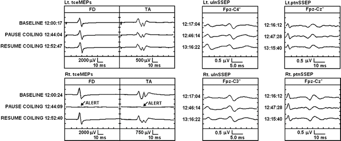
The fourth instance of vascular compromise occurred during coiling of an anterior communicating artery aneurysm. Following placement of several coils, there was abrupt loss of tceMEPs from the right upper and lower extremities, as shown in Fig. 42.2. Responses from the left upper and lower extremities were unchanged from baseline, as were cortical SSEPs to interleaved stimulation of the left and right ulnar nerves, respectively. Cortical SSEPs to stimulation of the right posterior tibial nerve showed only a nominal, clinically insignificant 25 % attenuation from baseline, while those to stimulation on the left side were unchanged. Placement of additional coils was halted temporarily and an arteriogram performed. The arteriogram showed no embolic occlusion or evidence of obvious vasospasm at that time; however, it is not implausible that this acute right upper and lower myotomal tceMEP loss represented an early functional manifestation of developing vasospasm not yet appreciated on angiography. Following a brief surgical pause, tceMEPs on the right side reappeared and response amplitudes returned to baseline range. Placement of coils subsequently was resumed and completed successfully with no additional episodes of tceMEP amplitude attenuation.


Fig. 42.2
Representative tceMEPs, ulnar nerve SSEPs, and posterior tibial nerve SSEPs recorded during neuroendovascular surgery for coiling of anterior communicating artery aneurysm. The top set of traces in each panel shows baseline-evoked potentials recorded during the early stages of the procedure. The middle set of traces shows evoked potentials following placement of several coils. Arrows indicate disappearance of tceMEPs from right first dorsal interosseous and tibialis anterior muscles at this time. The lower set of traces in each panel shows full recovery of previously attenuated potentials following a surgical pause and angiography to rule out vascular occlusion
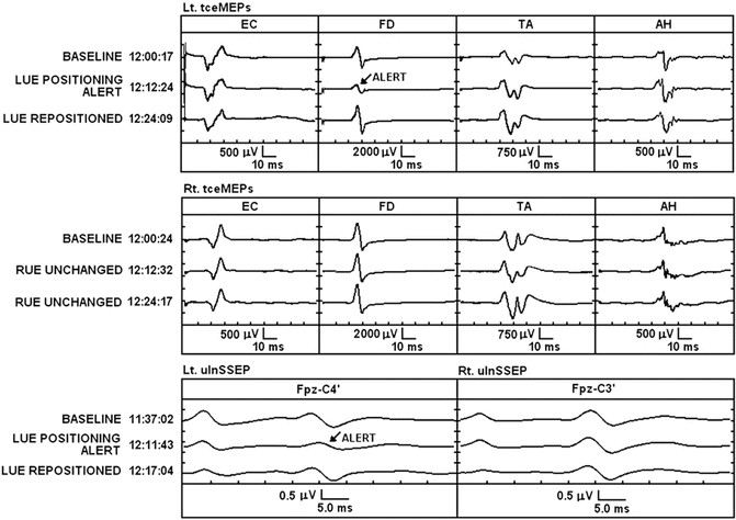
Upper extremity positioning alerts occurred in five (3.7 %) of 135 procedures and typically involved compression of the ulnar nerve. In each case, repositioning of the affected arm resulted in prompt resolution of neurophysiologic changes, as shown in Fig. 42.3. During the early stages of the procedure, tceMEPs from left first dorsal interosseous (FD) muscle and cortical SSEPs to stimulation of the left ulnar nerve decreased in amplitude by greater than 60 % from baseline. At this time, there were no concomitant changes in any of the other tceMEPs, including those from left extensor carpi radialis muscle, which unlike the FD muscle is innervated by the radial and not the ulnar nerve.


Fig. 42.3
Representative tceMEPs, ulnar nerve SSEPs, and posterior tibial nerve SSEPs recorded during the early stages of neuroendovascular surgery for coiling of anterior communicating artery aneurysm. The top set of traces in each panel shows baseline-evoked potentials. The middle set of traces shows evoked potential changes secondary to positioning of the left upper extremity. Arrows indicate attenuation of the tceMEPs from left first dorsal interosseous muscle and the cortical SSEP to stimulation of the left ulnar nerve. The lower set of traces in each panel shows full recovery of previously attenuated potentials following repositioning of the left upper extremity. LUE left upper extremity, RUE right upper extremity
In light of motor and sensory signal changes that appeared restricted to the left ulnar nerve, attention was directed to inspection of the left upper extremity. Following repositioning of the left arm, both the SSEPs and tceMEPs recovered to baseline amplitude.
Conclusions
Neurophysiological monitoring is effective in detecting evolving neural compromise and prompting timely intervention during neuroendovascular surgery. In the reported series, incidences of cerebrovascular and positioning-related neurophysiological changes were 3 % and 3.7 %, respectively. These results suggest that while the primary focus of neuromonitoring during neuroendovascular procedures is detection and reversal of developing iatrogenic injury secondary to vascular compromise, its role in identification of reversible positioning-related peripheral nerve compression should be neither overlooked nor underestimated. Finally, the high specificity of neuromonitoring during cerebrovascular surgery [6] is of value in confirming adequacy of collateral cerebral perfusion when there is a question of partial vessel occlusion during neuroendovascular therapy.
Success of monitoring during neuroendovascular surgery depends not only on the skill of the neuromonitoring team in detecting significant neurophysiologic change but also on close cooperation between the surgical neurophysiologist, anesthesiologist, and endovascular specialist in identifying the cause of change and initiating appropriate treatment.
Questions
- 1.
Somatosensory-evoked potentials are sensitive to ischemic insult of
- (a)
Afferent fibers within the internal capsule
- (b)
Efferent fibers within the internal capsule
- (c)
Cortical neurons of the post-central gyrus
- (d)
(a) and (c)
- (e)
(a), (b) and (c)
- (a)
- 2.
Multichannel EEG is most sensitive to ischemic injury of
- (a)
Basal Ganglia
- (b)
Internal Capsule
- (c)
Thalamus
- (d)
Cerebral Cortex
- (e)
Cerebellum
- (a)
- 3.
Get Clinical Tree app for offline access
Brain stem auditory-evoked potentials facilitate functional assessment of structures fed by the


