Chapter 82 Inotropes and vasopressors
The pharmacological support of the failing circulation is a fundamental part of critical care. The principal aim of these drugs is to restore inadequate systemic and regional perfusion to physiological levels.
DEFINITIONS
Inotropic agents are defined as drugs that act on the heart by increasing the velocity and force of myocardial fibre shortening. The consequent increase in contractility results in increased cardiac output and blood pressure. Characteristics of the ideal inotrope are shown in Table 82.1.
Vasopressors are drugs that have a predominantly vasoconstrictive action on the peripheral vasculature, both arterial and venous. These drugs are used primarily to increase mean arterial pressure.
The distinction between these two groups of drugs is often confusing. Many of the commonly used agents such as the catecholamines have both inotropic and variable effects on the peripheral vasculature that include venoconstriction, arteriolar vasodilatation and constriction.
Vasoregulatory agents may modulate the responsiveness of the peripheral vasculature to vasoactive drugs in pathological states such as sepsis. These agents include vasopressin and corticosteroids.
Given the overlap of pharmacodynamic effects of these drugs, the term ‘vasoactive therapy’ is a more appropriate description.
THE FAILING CIRCULATION
PHYSIOLOGY
Traditionally, cardiac output is discussed in terms of factors that govern cardiac function. These include preload, afterload, heart rate and rhythm, and contractility. Whilst this perspective is helpful in managing patients whose circulatory function is limited by cardiac disease, it is incomplete.
Cardiac output is controlled by the peripheral vasculature that is as energetic at returning blood to the heart as the heart is at pumping blood to the periphery1 (Figure 82.1).
Blood is pumped down a pressure gradient that is determined by the force of myocardial ejection (contractility) and impedance to ventricular ejection (afterload). The resultant mean arterial pressure is the major ‘afferent’ determinant of regional perfusion pressure. Twenty per cent of the blood volume is contained in the arterial (‘conducting’) vessels. There is a marked drop in perfusion pressure and flow across the capillary beds to allow diffusion of substrates and oxygen. The difference between mean arterial pressure and the pressure in end capillaries (‘efferent’ perfusion pressure) determines regional, or organ-specific, perfusion pressure.
Blood enters the venous system and is returned to the heart via a pressure gradient determined by mean systemic pressure and right atrial pressure. The amount of blood returned to the heart determines the degree of ventricular filling prior to systole (preload), which subsequently determines stroke volume and cardiac output.
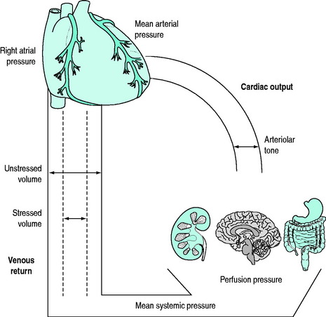
Under physiological conditions, the venous (‘capacitance’) system contains approximately 70% of the total blood volume which acts as a physiological reservoir (‘unstressed’ volume). Under conditions where circulatory demands increase, increased sympathetic tone will cause contraction of this reservoir. The resultant autotransfusion (‘stressed’ volume) may increase venous return by approximately 30% and subsequently cardiac output.2,3
Both the arterial and venous systems are integrated under complex neurohormonal influences. These include the adrenergic, renin–angiotensin–aldosterone, vasopressinergic and glucocorticoid systems in addition to local mediators such as nitric oxide, endothelin, endorphins and the eicosanoids.4
PATHOPHYSIOLOGY
Circulatory dysfunction or failure may be considered in terms of the major determinants of cardiac output, although there is marked interdependence between these factors.
HEART RATE FAILURE
Profound bradycardia will reduce both cardiac output and mean arterial pressure if sympathetic tone is compromised. Inotropes will increase both rate and speed of conduction, in addition to augmenting peripheral venous return, thereby restoring cardiac output and mean arterial pressure.
Tachycardia is associated with decreased left coronary artery perfusion, due to reduction of diastolic time, during which coronary perfusion occurs. This may exacerbate myocardial ischaemia in patients with coronary artery disease, particularly if mean arterial pressure, specifically diastolic blood pressure, is compromised. Therefore, drugs that shorten diastolic time or compromise coronary perfusion should be used with caution in susceptible patients.
PRELOAD FAILURE
Loss of intravascular blood volume or extracellular fluid is the most common cause of inadequate ventricular preload. This is corrected with appropriate fluids to maintain and restore a euvolaemic state. Hypovolaemia must be recognised and treated as soon as possible, preferably before vasoactive therapy is used.
There are other determinants of ventricular preload and venous return. Factors such as loss of muscle tone, positive intrathoracic pressure, loss of atrial systole (atrial fibrillation) and ablation of sympathetic tone will also compromise preload by reducing venous return. Under these circumstances, volume replacement alone may be insufficient to maintain adequate preload and vasoactive therapy may be required to increase venous return.
MYOCARDIAL FAILURE
Myocardial or ‘pump’ failure may be divided into disorders of systolic ejection (systolic dysfunction) and diastolic filling (diastolic dysfunction).
Systolic dysfunction occurs as a result of reduced effective myocardial contractility. This may be due to primary myocardial factors such as ischaemia, infarction or cardiomyopathy. Myocardial depression of both right and left ventricular function may occur in severe sepsis or following prolonged infusions of catecholamines. Increased impedance to ventricular ejection (e.g. hypertensive states) or structural abnormalities (e.g. aortic stenosis or hypertrophic obstructive cardiomyopathy) may cause systolic dysfunction.
Diastolic dysfunction is characterised by reduced ventricular compliance or increased resistance to ventricular filling during diastole. It may be due to mechanical factors such as structural abnormalities of the ventricle (e.g. restrictive cardiomyopathy) or to impaired diastolic relaxation that occurs with myocardial ischaemia or severe sepsis. This results in elevated end-diastolic pressure and pulmonary venous congestion. Episodic or ‘flash’ pulmonary oedema is a common clinical sign of diastolic dysfunction.5 Tachycardias that shorten diastolic time may exacerbate diastolic failure. Diastolic dysfunction frequently accompanies systolic failure, in both acute and chronic cardiac failure, particularly in elderly patients.
VASOREGULATORY FAILURE
Disruption or impairment of regulation of the peripheral vasculature may result in circulatory failure. This includes acute sympathetic denervation, such as high quadriplegia, epidural or total spinal anaesthesia (‘spinal’ shock); distributive failure such as anaphylaxis; or pathological ‘vasoplegia’ that occurs in severe sepsis.
These syndromes are characterised by reduced responsiveness of the peripheral circulation to endogenous or exogenous sympathetic stimulation. This results in pooling in the venous circulation due to the inability to provide a ‘stressed’ volume.4
Management of these conditions has traditionally focused on the arterial circulation with attempts to increase systemic vascular resistance, often regarded inaccurately as treatment of ‘afterload failure’. This is a misnomer as the problem is predominantly impaired venous return, compounded to a lesser extent by pathological arteriolar vasodilatation.6 Clearly, the effects of vasoregulatory failure will be exacerbated by concomitant hypovolaemia. Fluid loading to restore effective intravascular volume is essential.
CLASSIFICATION
The common ultimate cellular mechanism of action of these agents involves an influence on the release, utilisation or sequestration of intracellular calcium (Figure 82.2). These agents are divided into two main groups based on whether or not their actions depend upon increases in intracellular cyclic adenosine 3,5-monophosphate (cAMP) and are outlined in Table 82.2.

Figure 82.2 Schematic representation of the action of inotropic drugs on intracellular calcium in myocytes. AMP, adenosine monophosphate; ATP, adenosine triphosphate; cAMP, cyclic AMP; GR, glucagon receptor; Gs, G protein complex; IP, inositol phosphate 3; PDE III, phosphodiesterase III; PIP2, phosphoinositol diphosphate.
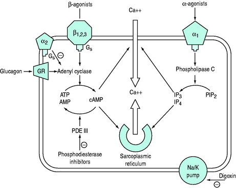
Table 82.2 Classification of inotropes
cAMP dependent | cAMP independent |
---|---|
Catecholamines (β-adrenergic agonists) | Catecholamines (α-adrenergic agonists) |
Adrenaline | Adrenaline |
Noradrenaline | Noradrenaline |
Dopamine | Dopamine |
Dobutamine | Digoxin |
Dopexamine | Calcium salts |
Isoprenaline | Thyroid hormone |
Phosphodiesterase inhibitors | |
Amrinone | |
Milrinone | |
Enoximone | |
Levosimendan | |
Calcium sensitisers | |
Levosimendan | |
Glucagon |
CATECHOLAMINES
Sympathomimetic amines are the most frequently used vasoactive agents in the intensive care unit (ICU) and include the naturally occurring catecholamines dopamine, noradrenaline (norepinephrine) and adrenaline (epinephrine), and the synthetic substances dobutamine, isoprenaline and dopexamine.
RECEPTOR BIOLOGY
PHYSIOLOGICAL
Agonists bind to populations of adrenergic receptors, largely divided into α and β subgroups. Further subgroups of α- (α1A, α1B, α2A, α2B, α2C) and β-receptors (β1, β2, β3) have been identified.7
β-Receptor occupancy predominantly activates adenyl cyclase to increase the conversion of adenosine triphosphate to cAMP. α-Receptor occupancy acts independently of cAMP by activation of phospholipase C which increases inositol phosphates (IP3 and IP4) and diacyl glycerol.
This complex agonist–receptor–effector relationship is responsible for homeostatic mechanisms such as physiological responses to stress and autoregulation.
PATHOPHYSIOLOGICAL
The activity and function of this system is dynamic and may be markedly influenced by pathological states. This mayresult in qualitative changes in the agonist–receptor–effector organ relationship (desensitisation) where receptors no longer respond to physiological or pharmacological sympathetic stimulation to the same extent. Quantitative changes such as reduced receptor density, receptor sequestration and enzymatic uncoupling (downregulation) may also result in impaired responses.8
BIOSYNTHESIS
The biosynthesis and chemical structures of the naturally occurring catecholamines are shown in Figure 82.3a.


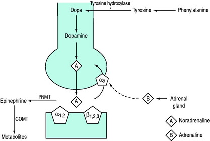
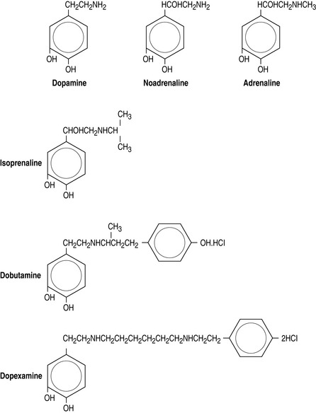
Figure 82.3 (a) Biosynthesis of catecholamines in sympathetic terminals. *Rate-limiting step by tyrosine hydroxylase. COMT, catechol-O-methyl transferase; PNMT, phenylethanolamine-N-methyltransferase. (b) Chemical structure of endogenous and synthetic catecholamines.
Catecholamines consist of an aromatic ring attached to a terminal amine by a carbon chain. The configuration of each drug is important for determining affinity to respective receptors.
Dopamine is hydroxylated to form noradrenaline, which is the predominant peripheral sympathetic chemotransmitter in humans, acting at all adrenergic receptors. The release of noradrenaline from sympathetic terminals is controlled by reuptake mechanisms mediated via α2-receptors and augmented by adrenaline released from the adrenal gland at times of stress. Noradrenaline is converted to form adrenaline that is subsequently metabolised in liver and lung.
All catecholamines have very short biological half-lives (1–2 minutes) and a steady state plasma concentration is achieved within 5–10 minutes after the start of a constant infusion. This allows rapid titration of drug to a clinical end-point such as mean arterial pressure.
Adrenaline and noradrenaline infusions produce blood concentrations similar to those produced endogenously in shock states, whereas dopamine infusions produce much higher concentrations than those naturally encountered. Dopamine may exert much of its effect by being converted to noradrenaline, thus bypassing the rate-limiting (tyrosine hydroxylase) step in catecholamine synthesis.
The synthetic catecholamines are derivatives of dopamine (Figure 82.3b). These agents are characterised by increased length of the carbon chain, which confers affinity for β-receptors. Dobutamine is a synthetic derivative of isoprenaline. These agents have relatively little affinity for α-receptors due to the configuration of the terminal amine, which differs from the endogenous catecholamines.
SYSTEMIC EFFECTS
The systemic effects of any of these agents will vary greatly between patients and within individuals at different times. Adequacy of response is often unpredictable and depends on the aetiology of circulatory failure and systemic comorbidities. In some patients, dramatic responses to small doses may occur, whilst in others, large doses of inotropes may be required to support the failing circulation.
The classification of sympathomimetic agents into α- and β-agonists, based on the above structure/function relationships, is only a crude predictor of systemic effects.
Adrenaline, noradrenaline and dopamine are all predominantly β-agonists at low doses, with increasing α-effects becoming evident as the dose is increased.
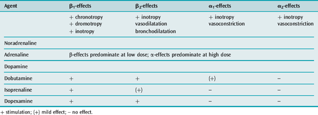
The synthetic catecholamines are all predominantly β-agonists.
CARDIOVASCULAR
The cardiovascular effects of the catecholamines under physiological conditions are shown in Table 82.3.
Noradrenaline, adrenaline and dopamine all tend to increase stroke volume, cardiac output and mean arterial pressure, with little change in heart rate and a low incidence of dysrhythmias. The effects on the peripheral vasculature are similar, with all agents increasing venous return without significant changes in calculated systemic vascular resistance.
Isoprenaline increases cardiac output predominantly by increasing heart rate and by moderate inotropy. This occurs without a significant change in blood pressure due to predominant β2-receptor induced veno- and vasodilatation.
The profile of dobutamine is similar to isoprenaline, although increases in heart rate are not as pronounced. Both of these agents may decrease mean arterial pressure, particularly in hypovolaemic patients, due to reduced venous return caused by venodilatation. The adverse effects of dobutamine and isoprenaline on heart rate and mean arterial pressure may compromise patients with ischaemic heart disease. However, the vasodilatory effects of dobutamine may be useful in selected patients with predominant systolic heart failure as a means of reducing afterload.
In the failing myocardium, particularly in patients with cardiac failure following cardiopulmonary bypass or septic shock, endogenous stores of noradrenaline are markedly reduced.9 Furthermore, there may be significant desensitisation and downregulation of cardiac β-receptors. In these situations, α1– and α2-receptors have an important role in maintaining inotropy and peripheral vasoresponsiveness.10 This may be expressed clinically as ‘tolerance’ or tachyphylaxis to catecholamines, particularly with predominantly β-agonists such as dobutamine. This phenomenon may explain the requirement for high doses of catecholamines in refractory shock states. Consequently, the role of β-agonists in patients with severe myocardial failure has been questioned.
Catecholamines have a significant effect on the venous circulation. These drugs primarily restore or maintain ‘stressed volumes’ of the capacitance vessels under pathological conditions, thereby maintaining or increasing cardiac output and mean arterial pressure. This is important in ‘vasoplegic’ states such as septic shock.11
In clinically used doses, intravenously administered catecholamines have minimal direct vasoconstrictive effects on conducting arterial vessels. Consequently, derived indices such as systemic vascular resistance do not reliably reflect the effect of catecholamines on the peripheral vasculature.
The development of peripheral gangrene in refractory septic shock has been attributed to catecholamine-induced vasoconstriction. There is little evidence to support this as the development of tissue gangrene in these situations primarily occurs as a consequence of intravascular thrombosis caused by sepsis-mediated coagulopathy.

Stay updated, free articles. Join our Telegram channel

Full access? Get Clinical Tree

