CHAPTER 35 Inguinal field block
Clinical anatomy
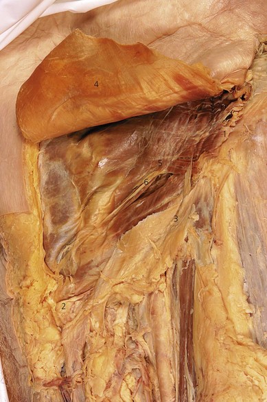
The iliohypogastric nerve is situated cephalad to the ilioinguinal nerve. At the level of the iliac crest, the iliohypogastric nerve divides into two terminal branches, the lateral cutaneous branch and medial cutaneous branches. The lateral cutaneous branch perforates the internal and external oblique and supplies the skin over the ventral part of the buttocks. This innervated area is behind that innervated by the subcostal nerve. The medial cutaneous branch continues ventrally until it pierces the internal oblique muscle above the anterior superior iliac spine, slopes downward between the internal oblique and external oblique muscles (Fig. 35.1), then pierces the external oblique aponeurosis 3 cm above the superficial inguinal ring, and ends by innervating skin over the lower part of the rectus abdominis and front of the pubis.
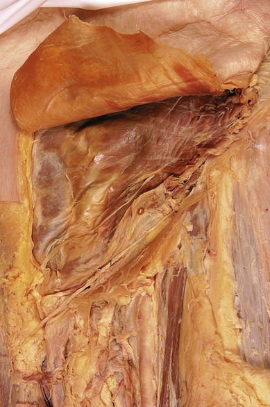
The ilioinguinal nerve runs ventrally, caudad to, and in a deeper plane than the iliohypogastric nerve. It perforates the transversus abdominis at the level of the anterior superior iliac spine and continues ventrally deep to the internal oblique (Fig. 35.2). Gradually, it pierces both internal and external oblique to reach the lower border of either the spermatic cord (in males) or the round ligament of the uterus (in females), where it finally reaches the inguinal canal. It contributes fibers to the internal oblique, the skin of the upper medial part of the thigh, and either the skin of the upper part of the scrotum and the root of the penis or the skin covering the labium majus and the mons pubis.
Ilioinguinal/iliohypogastric nerve block
Surface anatomy
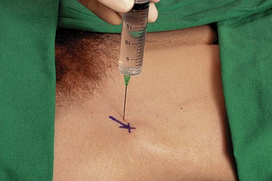
The important bony structure for the ilioinguinal/iliohypogastric nerve block is the anterior superior iliac spine. The needle insertion site for ilioinguinal and iliohypogastric nerve blocks is 1 cm medial and 1 cm inferior to the anterior superior iliac spine (i.e. above the inguinal ligament) (Fig. 35.3).
Sonoanatomy
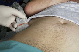
Perform a systematic anatomical survey from the iliac crest to the lower abdomen. The abdominal wall is scanned about 5 cm cranial to the anterior superior iliac spine. A sagittal oblique transducer orientation is used (Fig. 35.4). At this point, all three muscle layers of the abdominal wall can easily be identified by ultrasound and facilitate orientation (Fig. 35.5). The peritoneum and bowel are seen deeper to these (Fig. 35.5). The nerves appear as hypoechoic fascicular structures with hyperechoic rims sandwiched between the layers of muscle (Fig. 35.6). Trace the course of the nerves from above ASIS and distally towards the inguinal region. The iliohypogastric and ilioinguinal nerves consistently lie between the internal oblique and transversus abdominis muscles here. The recommended injection site for landmark-based approaches is situated medial to the anterior superior iliac spine. At this site, both nerves are often penetrating the internal oblique muscle. Performing a ‘blind’ technique here may result in difficulty for the injected local anesthetic to reach both nerves if they are not lying in the same compartment. This is a possible explanation for the high failure rates of 20–30%. It is more likely to reach both nerves with local anesthetic using the landmark-based approach where the nerves are lying in the same layer of the abdominal wall. Small vessels are frequently seen to accompany nerves within the plane.
< div class='tao-gold-member'>
Related posts:


Stay updated, free articles. Join our Telegram channel

Full access? Get Clinical Tree


