Fig. 13.1.
Anteroposterior (AP) view of the pelvis is acquired with internal rotation of the legs, laying out the femoral necks to evaluate for possible fracture. There is severe osteoarthritic change about the right hip with preferential loss of the superior, weight-bearing portion of the joint space (black arrow) and osteophytosis (curved black arrow) and relative preservation of the medial space. In contrast, the left hip is normal in appearance (white arrow). Chronic superior and inferior pubic rami fractures are seen on the right (thick black arrows) with interruption of the smooth cortical line and callus formation. Significant degenerative changes of the lumbar spine are partially visualized (asterisk).
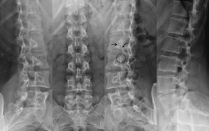
Fig. 13.2.
A four view series of the lumbar spine combines the traditional two view series (anteroposterior and lateral) with left and right oblique views. While the AP and lateral views are sufficient for evaluating for vertebral body height and alignment, oblique views reveal the “scotty dog” appearance of the posterior structures, allowing for evaluation of the pedicle (arrow), lamina (asterisk), pars articularis (curved arrow), and facets (circle). Spondylolysis, or interruption of the pars articularis, may predispose to neuroforaminal stenosis and subsequent neurogenic groin pain.
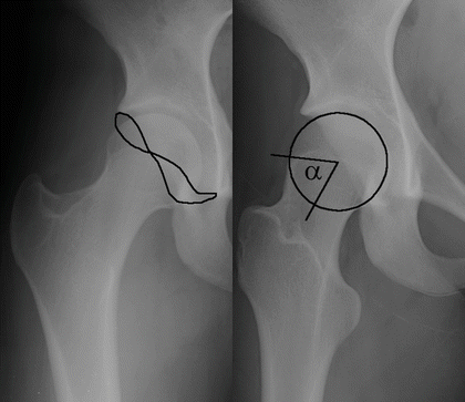
Fig. 13.3.
Two views of the right hip demonstrating the findings of mixed-type femoroacetabular impingement. Pincer-type deformity can be diagnosed on the anteroposterior view if there is evidence of acetabular overcoverage: in this case the anterior acetabular wall is somewhat more lateral than the posterior wall, forming a figure-of-eight. The modified Dunn view (patient lying supine with feet flat on the table) allows for the evaluation of cam-type deformity, which is due to asphericity of the femoral head. A circle is drawn within the confines of the femoral head and an alpha angle measured between the axis of the femoral neck and the point where the cortex of the neck first meets the head. This alpha angle measured 65°, while normal is considered less than or equal to 50°.
CT relies on the same principles as plain film radiography . Instead of acquiring a single image with a set amount of radiation, a CT scanner acquires many images from many angles, each of which requires much less radiation than a single conventional radiograph because they are not meant to stand alone. A sophisticated processing algorithm then integrates these individual “projections” into a three-dimensional attenuation map that can be viewed in the axial coronal or sagittal plane, or some oblique combination thereof. The advance of technology is such that current generation CT scanners can produce diagnostic images in any conceivable plane without appreciable loss of resolution. Pathologies such as appendicitis and degenerative spine disease in particular benefit from modern high-resolution multiplanar imaging (Fig. 13.4). Other less common causes of groin pain such as diverticulitis, abdominal aortic aneurysm, myositis ossificans, adductor tendonitis, prostatitis, and pelvic inflammatory disease are well visualized by computed tomography, although full characterization may require follow-up evaluation with contrast or another imaging modality.
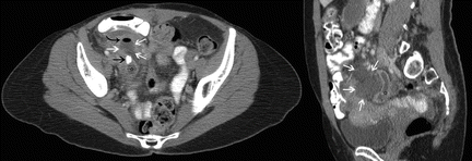

Fig. 13.4.
Axial and coronal CT images displaying a case of perforated appendicitis. The tubular shape of the appendix (asterisks) is clearly seen in the coronal view with a high-density fecalith (black arrow) best seen on the axial view. Gas (curved black arrow) within the surrounding fluid collection (surrounded by white arrows) is consistent with peri-appendiceal abscess formation.
The overall radiation dose of a CT study is a function of the dose associated with each acquisition, and the total number of acquisitions needed to generate the three-dimensional attenuation map. Patient factors such as body width, lean muscle mass, and the presence of intervening hardware (e.g., spinal fusion, hip arthrodesis, etc.) influence total dose. Modern technologies such as exposure control and iterative reconstruction can automatically reduce dose to minimally necessary levels. For comparison’s sake, the American College of Radiology (ACR) maintains a set of Appropriateness Criteria. Their most recent Radiation Dose Assessment lists a typical abdominal CT as the rough equivalent of ten single view radiographs [1]. This is in comparison to natural background levels of radiation, which averages an equivalent of about three radiographs per year in the United States [2]. The significant drop in radiation associated with CT has allowed for the application of dynamic acquisition in some diagnostic circumstances.
Intravenous iodinated contrast material has also advanced substantially since its early uses. Recent research suggests that the nonionic, low osmolarity contrast formulations currently employed in CT are not a causative factor in the development of nephropathy, particularly among patients with normal renal function at baseline [3, 4]. Rather, baseline glomerular filtration rate appears to be the primary determinant of acute kidney injury. Current iodinated contrast materials have been found to not represent an independent risk factor for AKI even among patients with impaired GFR (below 30 mL/min/1.73 m2) [5]. It bears mentioning that this research is relatively recent and requires independent confirmation before current practice guidelines will change [6]. The suggestion, however, is that the administration of iodinated contrast material should not be avoided in otherwise healthy individuals. While intravenous contrast material is not required for most protocols, it increases the ability of CT to evaluate for all manner of infectious, inflammatory, and neoplastic processes (Figs. 13.5, 13.6, and 13.7). Likewise, abdominopelvic evaluation by CT benefits substantially from routine oral contrast administration (Fig. 13.8). While rectal contrast agents also have utility, improvements in image resolution and multiplanar reformatting are leading to decreased reliance on such invasive administration.


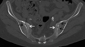


Fig. 13.5.
Axial, sagittal, and coronal CT images with features of delayed onset muscle soreness (DOMS), a form of exercise-induced muscular pain that, when involving the abdominal wall or pelvic musculature, may result in groin pain. With severe exertion, rhabdomyolysis and subsequent acute renal failure may occur. The soft tissues and fascial planes of the abdominal wall are diffusely edematous (white arrows) as compared to the unaffected tissues (curved white arrow) found more superiorly. Findings must be differentiated from fasciitis on the basis of clinical presentation and laboratory results.

Fig. 13.6.
Axial and coronal CT images of Fournier’s necrotizing fasciitis. Diffuse abdominal wall edema is again seen (white arrows), primarily involving the scrotum. Although subcutaneous air is sensitive for fasciitis, as in this case it is not always present. In contrast to prior patient, the clinical presentation here was that of sepsis secondary to pelvic infection.

Fig. 13.7.
Axial CT of the pelvis demonstrates bony erosions of the bilateral iliac wings (black arrows) with sparing of the sacrum (white arrows), consistent with sacroiliitis. Ferguson view of the pelvis (“pelvic outlet radiograph” not shown) will accentuate the sacroiliac joints and may reveal sacroiliitis without the need for CT.

Fig. 13.8.
Axial, sagittal, and coronal CT reveal loop of bowel (asterisks) exiting the peritoneal cavity below the inguinal ligament and medial to the femoral vessels (black arrows), diagnostic of femoral hernia.
CT contrast reactions are infrequent occurrences that are incompletely understood. In the majority of patients, reactions to iodinated contrast are not allergies in the traditional sense of IgE mediation, yet may present with anaphylactoid airway edema or other severe physiologic consequences all the same [7]. As per the above discussion of contrast-induced nephropathy, the incidence of severe adverse reactions has decreased substantially since the switch was made to low osmolarity contrast material [8]. A history of asthma or prior contrast reaction increases the risk of acute reaction. Pretreatment with oral corticosteroids is recommended for at-risk patients [9]. Common pretreatment regimens for high-risk individuals may involve oral administration of 50 mg prednisone at 13, 7, and 1 h before contrast administration, with or without oral administration of 50 mg diphenhydramine and 25 mg ephedrine at 1 h before contrast administration [10], though facility-specific variations abound. Recent studies suggest that multiple exposures to contrast material may be necessary for severe reaction to occur [11].
Magnetic Resonance
Magnetic resonance scanners utilize low-energy light to interact with the hydrogen atoms found throughout most organic tissues. The electromagnet inside an MR scanner is always “on,” and typically operating at 1.5 T of field strength (roughly 10,000 times the strength of the Earth’s natural field) although 3 T scanners are becoming more widely available in routine clinical imaging. This magnetic field provides energy to hydrogen atoms, forcing them to line up along the direction of the magnet much in the way that a compass needle will line up with the Earth. Once the hydrogen atoms line up, the machine can communicate with them by sending out radio-frequency pulses that only specific atoms are able to respond to, forcing them to change direction and oppose the magnetic field. This opposition is relatively unstable and the flipped hydrogen atoms will eventually switch back to their natively aligned state, emitting their own radio-frequency pulse and communicating back with the scanner as they do so. Since every tissue is different, the rate of this process varies dramatically throughout the body and allows for tissue discrimination. Foreknowledge of how fat, water, and other body substances will behave under these conditions allows for the targeting by specific “sequences” such as T1-weighted sequences for fat, T2-weighted sequences for water, and short–tau inversion recovery (STIR) for edema, among many others. Compare this to computed tomography, where there is only one parameter (i.e., density) that significantly impacts tissue discrimination.
MR excels at differentiating between many subtle soft tissue and musculoskeletal pathologies responsible for groin pain, such as those seen in iliopsoas tendinosis, bursitis, osteitis pubis, and athletic pubalgia (Figs. 13.9, 13.10, and 13.11). Avascular necrosis of the femoral head in particular is apparent on MR long before it is demonstrable by CT (Fig. 13.12). Yet MR is not always a definitive examination, particularly with respect to certain musculoskeletal pathologies in which the relative lack of hydrogen in bone can necessitate correlation with conventional radiography or CT. While radiographically occult stress fractures are clearly identified on MR, many other benign and malignant osseous lesions may be indistinguishable from each other on the basis of MR alone. Definitive evaluation of acetabular labral tears is via MR arthrography, wherein contrast material is directly injected into the hip joint (Fig. 13.13).
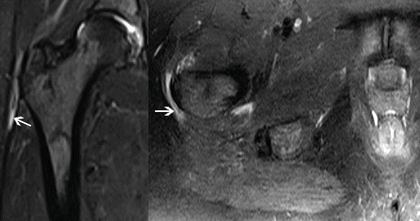

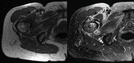
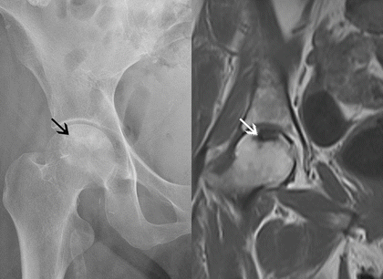

Fig. 13.9.
Coronal and axial T2-weighted MR through the pelvis show increased fluid signal (white arrows) lateral to the greater trochanter, consistent with greater trochanteric bursitis.

Fig. 13.10.
Coronal and axial CT of the pelvis demonstrate diffuse subchondral sclerosis (black arrows) of the pubic symphysis. Corresponding coronal and axial T1-weighted MR demonstrate focal hypointensity (white arrows), consistent with osteitis pubis.

Fig. 13.11.
Axial T1- and T2-weighted MR through the pelvis demonstrating significant edema (black arrow) and trace fluid within the adductor compartment, consistent with low-grade adductor strain.

Fig. 13.12.


Anteroposterior radiograph of the right hip reveals subtle sclerosis (black arrow) representative of avascular necrosis. Coronal T1-weighted MR demonstrates serpiginous hypointensity (white arrow), confirming the diagnosis.
Stay updated, free articles. Join our Telegram channel

Full access? Get Clinical Tree


