Ileus

24 Ileus


Ileus is defined as disruption of coordinated physiologic bowel motility owing to a nonmechanical cause.1 As a result, intestinal contents cannot progress through the gastrointestinal (GI) tract. The word ileus is derived from the Greek eileos, which means “twisting.” An ileus can develop as a primary process or as a result of a separate process that is usually associated with inflammation. The diagnosis of ileus must be differentiated from the diagnosis of mechanical bowel obstruction, since the latter condition also blocks the normal aboral progression of bowel contents but is due to the presence of an extrinsic or intrinsic anatomic barrier. These two conditions are treated differently.


image Pathophysiology


Physiologic bowel motility is a complex process that results from the interaction of various neural networks and neurohormonal mediators. During the fasting state, the coordinated contractions of the GI tract are referred to as migrating motor complexes (MMC). The contractions can be viewed as occurring in three phases: the resting phase, intermittent contractions of moderate amplitude, and high-pressure waves. When a food bolus is introduced into the intestine, the MMCs terminate, and the digested food, or chyme, is propelled through the GI tract via coordinated contractions of the smooth muscle in the intestinal wall, also referred to as peristalsis. This process is regulated primarily by the enteric nervous system (ENS), which is comprised of myenteric and submucosal sensory and motor nerve plexi and the interstitial cells of Cajal. The ENS transmits sensory information from the intestinal wall to the central nervous system (CNS) via a network of visceral sensory afferents in the vagus, splanchnic, and pelvic nerves. The ENS also connects the visceral motor efferents in these same nerves with the intestinal smooth muscle cells. The ENS and intestinal smooth muscle activity are inhibited by sympathetic signaling and stimulated by parasympathetic cholinergic signaling. Alternatively, the ENS can function independently of CNS control via the autonomic nervous system through secreted mediators that include substance P, vasoactive intestinal peptide, and nitric oxide.


Ileus can develop when physiologic neural signaling and neurohormonal networks are disrupted. Ileus can result from the presence of inhibitory neuroenteric signaling through increased sympathetic activity, inflammation of surrounding organs or the bowel wall itself, paracrine and endocrine activity of inhibitory gastrointestinal peptides or endogenous opioids, and the use of exogenous opioids for analgesia. The most common clinical situation associated with ileus is the immediate period following abdominal operations. In normal circumstances, physiologic small-bowel motility returns within the first 24 hours after the procedure, gastric motility returns within 24 to 48 hours, and colonic motility within 48 to 72 hours. If the return of normal GI function exceeds these time limits, or ileus develops that is independent of a recent operation, a cause for ileus should be sought.


image Clinical Features and Diagnosis


Most patients with ileus exhibit abdominal distension, poorly localized bloating and pain, inability to tolerate oral intake, nausea and vomiting, and obstipation. The absence of bowel sounds on abdominal examination can help distinguish ileus from mechanical bowel obstruction; in the latter condition, high-pitched bowel sounds and/or borborygmi are often audible. Patients with severe and advanced cases of ileus can present with peritonitis due to intestinal ischemia or perforation from bowel dilatation, as well as abdominal compartment syndrome.


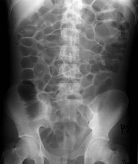
Radiographic studies are often obtained during the evaluation of patients with suspected ileus. Abdominal radiographs sometimes can be helpful for differentiating ileus from mechanical small bowel obstruction. The presence of gas in the stomach, small intestine, and colon (Figure 24-1) suggests ileus. In contrast, a paucity of gas within the abdomen, air/fluid levels within the small bowel, and absence of air within the colon suggest mechanical small bowel obstruction (Figure 24-2). A computed tomography (CT) scan with enteral contrast administration can better distinguish patients with ileus from those with mechanical bowel obstruction. Inspection of the abdominal CT scan often makes it possible to accurately localize a point of obstruction or a region of transition from dilated to decompressed bowel. If these findings are present, the diagnosis of mechanical bowel obstruction is established. Passage of oral contrast into the colon within 4 hours favors ileus over a bowel obstruction as the cause of intestinal dysmotility. The CT scan can also identify other intraabdominal inflammatory processes that can be the cause of ileus (e.g., appendicitis, pancreatitis, intraabdominal abscess).


< div class='tao-gold-member'>

Stay updated, free articles. Join our Telegram channel

Jul 7, 2016 | Posted by in CRITICAL CARE | Comments Off on Ileus

Full access? Get Clinical Tree

Get Clinical Tree app for offline access