Idiopathic and Thrombotic Thrombocytopenias

Chapter 63


Idiopathic and Thrombotic Thrombocytopenias


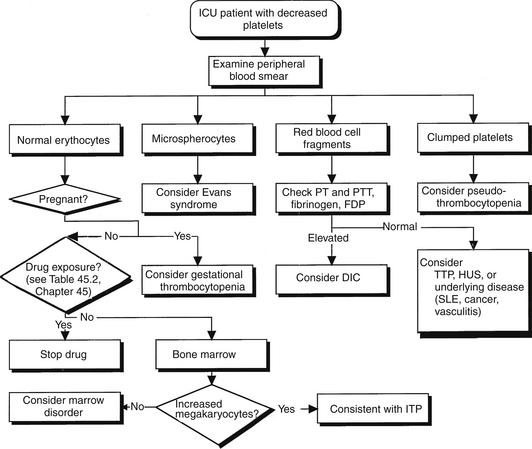
Moderate to severe thrombocytopenia is a major feature of several disorders that accompany or lead to stays in the intensive care unit (ICU). Immune (idiopathic) thrombocytopenic purpura (ITP) and thrombotic thrombocytopenic purpura (TTP) are two such conditions. Early and accurate diagnosis and rapid application of appropriate therapy are keys to the successful management of patients with these disorders. Accurate diagnosis requires differentiating the thrombocytopenia of ITP and TTP from each other as well as from other thrombocytopenic conditions. The diagnostic workup should begin with a carefully focused history and physical examination, followed by an examination of the peripheral blood smear (Figure 63.1). Blood products should not be given to a nonbleeding patient with thrombocytopenia until the smear is inspected, particularly to evaluate for the presence or absence of schistocytes and rule out the presence of TTP. Moreover, during the initial evaluation, patients should be safeguarded from iatrogenic bleeding complications resulting from a low platelet count (Box 63.1).


Immune Thrombocytopenic Purpura


Mechanism and Diagnosis


Autoantibodies directed against the patient’s platelets cause ITP. These antibodies are usually directed against the platelet glycoproteins Ib/IX or IIb/IIIa, and their binding results in platelets being destroyed in the spleen. Destruction occurs by splenic macrophage Fc-receptor recognition of antibody-coated platelets. An additional contributing factor to thrombocytopenia observed in ITP is decreased platelet production. Patients with ITP usually have mucosal bleeding, severe anemia, or incidental thrombocytopenia on routine blood counts. Fortunately, severe bleeding such as intracranial hemorrhage is rare in ITP.


ITP is characterized by a decrease in the number of platelets seen in the peripheral smear. Occasionally, large platelets can be found. Erythrocytes and leukocytes should appear normal. Bone marrow examination may show an increased number of megakaryocytes, indicating an appropriate response to the increased platelet destruction. Platelet antibody testing is usually unhelpful because of low sensitivity and specificity.


ITP remains a diagnosis of exclusion after other causes of thrombocytopenia are ruled out. Box 63.2 lists the differential diagnosis of thrombocytopenia without microangiopathy. Evans syndrome is also an antibody-mediated disease, with antibodies directed against both platelets and erythrocytes. The destruction of erythrocytes occurs with the same mechanism as does the destruction of platelets. Bone marrow aspiration is an appropriate test to establish the diagnosis in patients older than 60 or in those considering splenectomy.


ITP has been associated with other conditions in up to 20% of cases. In this setting, it is often called secondary ITP. Causes of secondary ITP include collagen vascular diseases, lymphoproliferative disorders, infections, and other autoimmune syndromes. Thrombocytopenia is also a common manifestation of human immunodeficiency virus (HIV) disease, presumably caused by increased platelet destruction on an immune basis. The clinical features of HIV-associated ITP are similar to those of idiopathic ITP. However, ITP associated with HIV infection in patients with hemophilia is of particular concern because these patients are at much higher risk for catastrophic bleeding than are patients with primary ITP.


Treatment


There are few well-controlled clinical studies on which to base recommendations for treatment of adult patients with ITP. Most of the literature on the treatment of ITP consists of case series without a control group. Treatment of ITP depends on the platelet count and clinical presentation. Spontaneous bleeding in ITP is rare with platelet counts > 50,000/μL. This number of platelets is also sufficient for most surgical procedures. Although rare, spontaneous bleeding has been reported in ITP patients with platelet counts in the 30,000 to 50,000/μL range. Because as many as 5% of adults and 40% of children with ITP have spontaneous remissions, it is safe to observe patients with ITP and platelet counts > 30,000/μL if they are not actively bleeding.


Actively bleeding patients with ITP require urgent management. Platelet transfusions are usually ineffective because of shortened platelet survival despite platelet counts increasing transiently in some patients. Pretreatment with intravenous immunoglobulin (IVIG) at a dose of 0.4 to 1.0 g/kg may increase the life span of the transfused platelets and cause an increase in the platelet count several days later by modulating the immune response. This therapeutic modality is expensive, costing between $5000 and $10,000 per treatment course. Antifibrinolytic agents such as epsilon-aminocaproic acid can effectively reduce hemorrhage, but the use of these agents is complicated by an increased incidence of thrombosis.


Glucocorticoids have become the mainstay of treatment for ITP that is symptomatic or when the platelet count falls to < 30,000/μL. The typical daily dose of prednisone is 1 mg/kg. This inexpensive treatment elevates platelet counts in about two thirds of treated patients. Most patients exhibit a decline in platelet count when the prednisone dose is tapered and therefore require additional therapy. The anti-CD20 monoclonal antibody, rituximab, has shown efficacy in patients who relapse following glucocorticoid therapy. Rituximab is typically given by monthly intravenous infusions of 375 mg/m2 for four doses. Alternatively, anti-D immune globulin has shown efficacy in patients with ITP who are Rh positive and have an intact spleen. Splenectomy can produce sustained increases in platelet counts in more than two thirds of patients with ITP. Pneumococcal, meningococcal, and hemophilus vaccines should be given 2 weeks before splenectomy to reduce the risk of severe complications resulting from postsplenectomy pneumococcal bacteremias. Some patients have a relapse of ITP months or even years after splenectomy. This is attributable to the presence of an accessory spleen. These spleens occur in up to 20% of patients and are easily detected by radionuclide scanning. After the removal of accessory spleens, ITP resolves in many of these patients.


ITP may become refractory to conventional therapy in some patients. There is no evidence and very little consensus about how to treat patients who do not respond or who respond transiently or incompletely to initial therapy with glucocorticoids. Modalities of therapy for refractory ITP include the use of immunosuppressive agents, such as azathioprine, cyclophosphamide, or vinca alkaloids. Alternatively, danazol, an androgenic steroid, can be useful for sustaining partial remissions or a decrease the glucocorticoid dose. Small case series report that combination chemotherapy and high-dose dexamethasone is efficacious in the management of chronic refractory ITP.


As mentioned previously, the monoclonal antibody against CD-20, rituximab, has been used in patients with refractory ITP, either before or after splenectomy. Again, most of the data here derive from case series and systematic reviews. The majority of these studies had a relatively short follow-up time, and based on this evidence, rituximab was suggested to be not as effective as splenectomy for establishing durable complete responses, nor was it safer than splenectomy.

< div class='tao-gold-member'>

Stay updated, free articles. Join our Telegram channel

Jul 7, 2016 | Posted by in CRITICAL CARE | Comments Off on Idiopathic and Thrombotic Thrombocytopenias

Full access? Get Clinical Tree

Get Clinical Tree app for offline access