 Clinical suspicion of hip dislocation
 Hip pain with obvious deformity in the setting of a motor vehicle crash, pedestrian struck by a vehicle, falls, or sports-related injuries
 Radiographic evidence of hip dislocation
CONTRAINDICATIONS
 Associated femoral neck fracture
 Coexistent fracture in dislocated extremity
RISKS/CONSENT ISSUES
 Inadvertently converting a dislocation to a fracture-dislocation (acetabulum or femoral head fracture)
 More common in the elderly with osteoporotic bones
 Oversedation may lead to inability to protect the airway with subsequent potential risk of aspiration
 General Basic Steps
 Obtain radiographs
 Sedation/Analgesia
 Have assistants for help
 Perform procedure
LANDMARKS
 Posterior Hip Dislocation
 Mechanism of injury—femoral head is forced out of the acetabulum and rests posteriorly
 Clinical features—affected extremity shortened, adducted, and internally rotated; patient may hold hip flexed with knee of affected extremity resting on opposite knee (FIGURE 66.1)
 Radiographic evidence—femoral head resting posterior to the acetabulum (FIGURE 66.2)
 Anterior Hip Dislocation
 Mechanism—forced abduction with the hip in a flexed position or forced hyperextension of the hip
 Clinical features—affected extremity abducted, slight flexion, and externally rotated
 Radiographic evidence—femoral head dislocated medially toward obturator foramen (obturator dislocation) and femoral head dislocated laterally toward pubis (pubic dislocation) (FIGURE 66.3)
TECHNIQUE
 Preprocedure
 Radiographs
 Should be obtained preprocedure only if there is a concern for a fracture or to determine the position of the dislocation
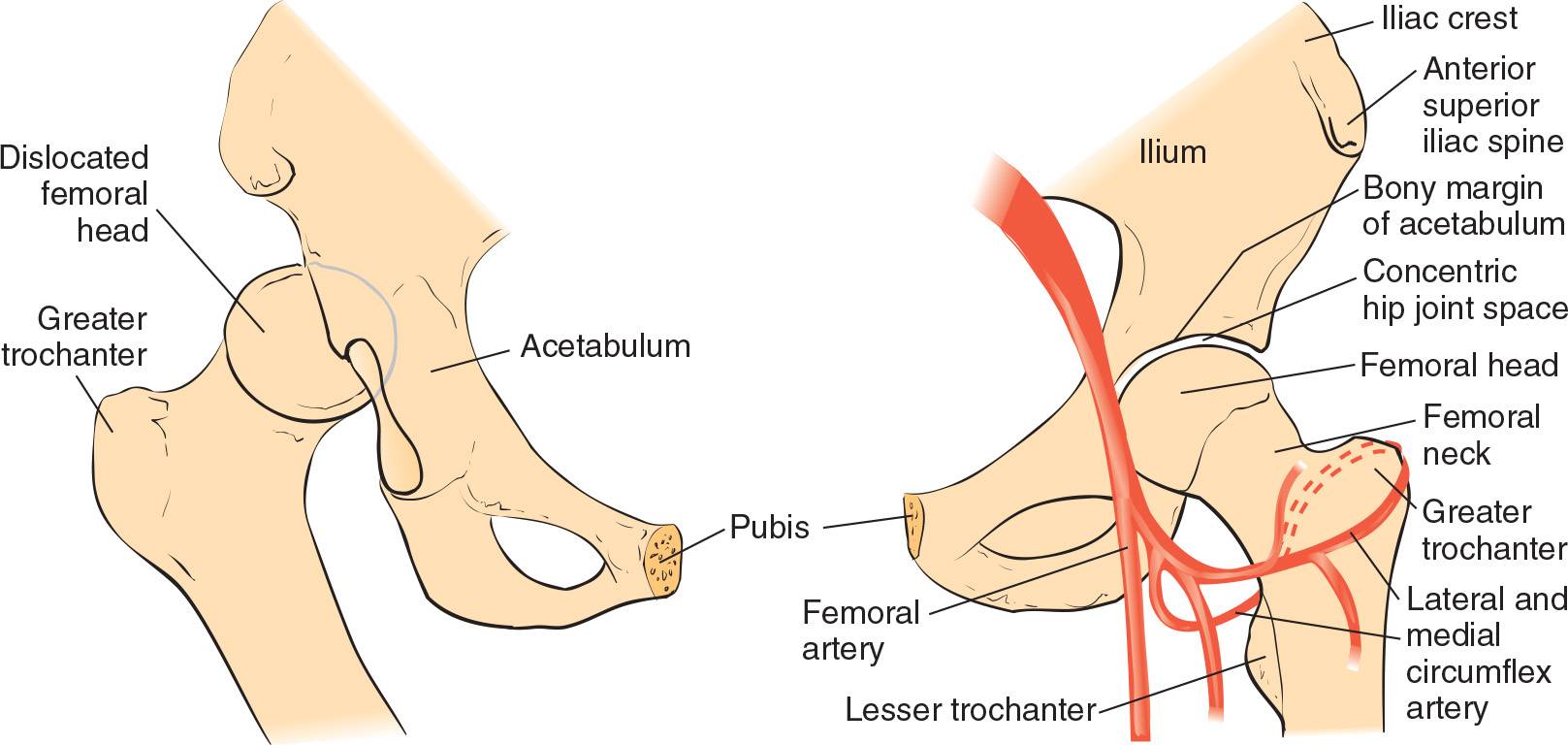
FIGURE 66.1 Normal (left) and dislocated (right) hip. (From Young GM. Reduction of common joint dislocations and subluxations. In: Henretig FM, King C, eds. Textbook of Pediatric Emergency Procedures. Philadelphia, PA: Williams & Wilkins; 1997:1093, with permission.)
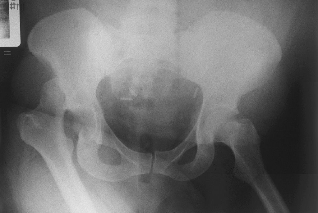
FIGURE 66.2 Anteroposterior pelvis radiograph of a posterior hip dislocation of the right hip. (From Tornetta Paul III. Hip dislocations and fractures of the femoral head. In: Bucholz RW, Heckman JD, Court-Brown C, eds. Rockwood and Green’s Fractures in Adults. Vol 2. 6th ed. Philadelphia, PA: Lippincott Williams & Wilkins; 2006:1718, with permission.)
 Posterior Dislocation Reduction
 Allis Maneuver (FIGURE 66.4)
 Patient is placed supine
 Downward stabilization of the pelvis is performed by an assistant
 With the knee flexed, apply traction in-line with the deformity with gentle flexion of the hip to 90 degrees
 Perform gentle internal-to-external rotation as the hip is flexed
 Once reduction is achieved, hip is brought to the extended position while traction is maintained
 Legs are then immobilized in slight abduction through the placement of pillows between the knees
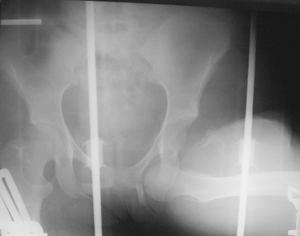
FIGURE 66.3 Anteroposterior pelvis radiograph of an anterior hip dislocation of the left hip. (From Tornetta Paul III. Hip dislocations and fractures of the femoral head. In: Bucholz RW, Heckman JD, Court-Brown C, eds. Rockwood and Green’s Fractures in Adults. Vol 2. 6th ed. Philadelphia, PA: Lippincott Williams & Wilkins; 2006:1720, with permission.)
Related posts:



Stay updated, free articles. Join our Telegram channel

Full access? Get Clinical Tree


