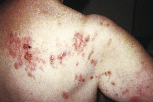
Glen Blair Herpes zoster (shingles) is a dermatologic eruption caused by reactivation of the varicella-zoster virus (VZV) that follows, sometimes by decades, a primary varicella-zoster (chickenpox) infection. A prodrome of pain or dysesthesia may precede by several days a vesicular eruption that typically occurs in a unilateral dermatomal distribution. Vesicular lesions appear during several days and may last for 7 to 10 days or more, although they can last for more than 4 weeks on rare occasions. The eruption can be extremely painful, and 5% to 20% of patients go on to develop a protracted pain syndrome called postherpetic neuralgia (PHN).1 One in three persons will develop zoster during their lifetime, roughly 1 million Americans per year.2 After primary infection, VZV lies dormant in the sensory root ganglion, kept in check by the host’s acquired cell-mediated immunity. The zoster eruption results from reactivation of the latent varicella infection in the dorsal root or cranial nerve ganglion cells, a process that is thought to be caused by an age-related decline in cell-mediated immunity.3 Zoster incidence is seen to increase with age and immune suppression. Zoster is common and usually self-limited in adults, but serious complications can occur that may require consultation. Involvement of the ophthalmic branch of the trigeminal nerve can result in ocular keratitis, scarring, and loss of vision. Patients who are immunocompromised may have a disseminated infection that can result in a diffuse varicella-like eruption, neurologic complications, or visceral involvement that can cause death, most commonly from pneumonia.2 After initial varicella infection, the virus lies dormant in the dorsal root ganglia. Once it is reactivated, the virus replicates and travels through the axons, spreading from cell to cell until it penetrates the epidermis. Reactivation is likely to be multifactorial, age being the most important risk factor. Immunosuppressed individuals of all types are at increased risk, as are some populations with chronic comorbid diseases, such as rheumatoid arthritis and inflammatory bowel disease. Trauma, surgery, and a recent cancer diagnosis have also been linked to zoster reactivation. Anecdotal references to stress as a cause have not been supported by research, but those with severe physical limitations as well as older women are certainly at higher risk.2 Herpes zoster lesions contain high concentrations of virus that can be spread by contact and by air but are less contagious than primary infection. Contagion is possible once the rash has appeared and continues until the lesions have crusted. Zoster classically is seen as a unilateral eruption within one dermatome; however, 20% of the time, an adjacent dermatome will be involved. The eruption is often preceded by a prodrome of pain, dysesthesia, or pruritus in the affected dermatome. Pain can be described as stabbing, burning, aching, or excruciating. The eruption is initially erythematous and maculopapular and becomes clusters of clear vesicles during the course of several hours (Fig. 55-1). New lesions may continue to develop for several days. Low-grade fever and lymphadenopathy may be present. The most common areas of involvement are the thoracic, cranial (especially the trigeminal), and lumbar nerves. On the rare occasion that there is no visible eruption, a condition called zoster sine herpete, the pain is sometimes attributed to other causes (e.g., angina, renal colic, pleural pain, sciatica, or migraine), depending on the dermatome affected. There is lack of consensus as to how long after rash resolution the pain needs to persist to be considered PHN. Regardless, pain can exist in varying degrees for many years. Diagnosis is based on the clinical presentation of a vesicular eruption in a unilateral, dermatomal distribution. Without the skin eruption, zoster is a diagnosis of exclusion. Disseminated herpes zoster is a generalized eruption of lesions along with the typical segmental distribution and occurs primarily in immunocompromised individuals. A Tzanck test is a rapid way to confirm the diagnosis of zoster in the provider’s office but does not distinguish between VZV and herpes simplex virus. The direct fluorescent antibody (DFA) test is another rapid test that is available in some settings. Polymerase chain reaction (PCR) analysis, although not available in all settings, is rapid and sensitive. All of these tests are preferred to viral culture because they are more rapid and often more sensitive.4 Diagnostics Herpes Zoster
Herpes Zoster (Shingles)
Definition and Epidemiology
Pathophysiology
Clinical Presentation and Physical Examination
Diagnostics
![]()
Stay updated, free articles. Join our Telegram channel

Full access? Get Clinical Tree


Herpes Zoster (Shingles)
Chapter 55