Head Blocks


Head Blocks


A. A.J. Maillard


Historically, the best-known major surgical procedure of the head performed under regional anesthesia in the United States is the partial maxillectomy performed on President Grover Cleveland in 1893. This chapter is presented according to the anatomic sites and types of nerves (scalp and face, facial nerves, trigeminal nerve, midface, and mandibular nerve). For each site, the relevant anatomy and the corresponding blocks will be described. Since most head blocks involve cranial nerves or their branches, consideration will also be given to the bony anatomy and especially the neural foramina. All of these blocks can be performed with the patient in the sitting or supine position depending on the block and the condition of the patient.


A. Blocks of the Scalp and Face

Indications: Blocks of the cutaneous nerves supplying the scalp are indicated in the emergency room and for plastic surgery to avoid tissue distortion. Blocks of the frontal branch of the supraorbital and infraorbital nerves are indicated for anesthesia in or around the eye. These blocks are indicated for blepharoplasty or for other lid procedures, including traumatic and posttraumatic reconstructive surgical procedures. Blocks of the auriculotemporal, greater auricular nerves, and minor occipital nerves are indicated for ear surgery.

Needle Size: 25-gauge, 20-mm needle.

Volume: 1 to 2 mL.

Anatomic Landmarks: Cutaneous nerves supplying the scalp and face (Fig. 23-1): 1, frontal nerve; 2, supraorbital nerve; 3, zygomaticotemporal nerve (V2); 4, auriculotemporal nerve (V3); 5, greater auricular nerve; 6, minor occipital nerve; 7, greater occipital nerve; 8, supra- and infratrochlear nerve; 9, infraorbital nerve; 10, external nasal branches of the ethmoid nerve; 11, mental nerve.

Approach and Technique: The appropriate site is identified, and 0.5 to 1.0 mL of 1% lidocaine with 1/100,000 epinephrine is injected subcutaneously in small increments up to 2 mL per nerve to obtain adequate anesthesia.

Tips


  • Originally, these blocks were directed at specific cutaneous nerves to block their sensory distribution. However, as epinephrine came into general use, its hemostatic effects on the scalp became a great aid to neurosurgeons. Therefore, instead of electively blocking these nerves, surgeons started to infiltrate the scalp around their proposed incision sites.


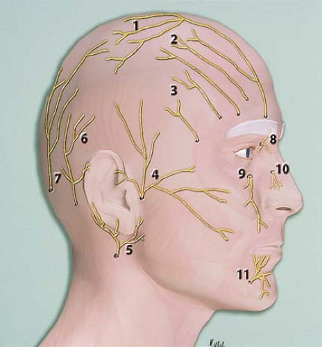
Figure 23-1. Cutaneous nerves supplying the scalp and face.


B. Blocks of the Facial Nerve

Indications: Blocks of the facial nerve are indicated when cosmetic procedures are planned that require the injection of botulin toxin-A to produce nerve paralysis with subsequent removal of forehead rhytides. It is of utmost importance to test the effect of blocking the zygomaticotemporal branch of the facial nerve using a short-acting local anesthetic. If the botulin injection is not performed correctly, and the botulin toxin is infiltrated lower, then a paralysis of the eyelids occurs that can last 2 to 3 months (temporary motor denervation). Blocks of the facial nerve are also used for plastic surgery (to produce a temporary paralysis of the eyelids when performing CO2 laser resurfacing or dermabrasion) and for traumatic repairs as well as posttraumatic reconstructive surgery.

Needle Size: 25-gauge, 35-mm needle.

Volume: Up to 2 mL.

Anatomic Landmarks: The orbit.

Approach and Technique: 1% lidocaine with 1/100,000 epinephrine is injected in small increments (up to 2 mL) along the orbit posterior to the rim, laterally and inferiorly. The needle can come close to the midline without causing paralysis and affecting eye closure. The effects are not long lasting and can be used as a good test prior to injecting the botulin toxin.

Tips


  • The combination of a supraorbital nerve block and an infraorbital nerve block provides complete anesthesia of the periorbita (Fig. 23-2).


  • The advantages of this technique over infiltrating techniques are that the volume of the soft tissues is not augmented by these injections and that there is no soft tissue distortion. Therefore, the surgeon has much better perspective to plan and carry out the repairs.


Figure 23-2. The combination of a supraorbital nerve block and an infraorbital nerve block provides complete anesthesia of the periorbita.


C. Blocks of the Trigeminal Nerve and Associated Dermatomes for V1, V2, and V3

The first division of the trigeminal nerve (V1) gives rise to the supraorbital nerve, which exits the brain through the supraorbital fissure. This branch gives sensation to the forehead and parietal scalp. The second division (V2) exits through the infraorbital fissure to exit into the face through the infraorbital foramen, located at approximately the midpoint of the infraorbital rim. Figure 23-3 depicts the cutaneous dermatomes associated with the different sensory branches of the trigeminal nerve.

Only gold members can continue reading. Log In or Register to continue

Stay updated, free articles. Join our Telegram channel

Jun 19, 2016 | Posted by in PAIN MEDICINE | Comments Off on Head Blocks

Full access? Get Clinical Tree

Get Clinical Tree app for offline access