Pulmonary physiology of aging.
In the cardiovascular system, decreased cardiac relaxation due to myocardial stiffening leads to impaired diastolic filling [15]. Vascular elasticity decreases, making hypertension and compensatory left ventricular hypertrophy more likely. Autonomic control is degraded, reducing responsiveness to β–adrenergic stimulation, increasing orthostatic hypotension, and impairing thermoregulation. Additionally, exercise tolerance decreases while arteriosclerosis, valvular problems, and arrhythmias increase [11]. These changes can result in fatal consequences. Myocardial infarction is the leading cause of postoperative death in octogenarians [12]. Further, patients older than 65 years who have had a previous MI have increased rates of reinfarction after non-cardiac procedures when compared with the general population [16].
Cardiac health in the geriatric population may be assessed preoperatively by estimating exercise tolerance associated with their daily activities. For a comprehensive overview, consult the American College of Cardiology and the American Heart Association 2014 published guidelines on cardiac risk in non-cardiac surgery [17–18].
Geriatric patients are disproportionately at risk for postoperative delirium (POD) and postoperative cognitive dysfunction (POCD). Delirium is an acute change in cognition, attention, or awareness with many different etiologies. Delirium is temporary, unlike dementia. Postoperative delirium is categorized as hyperactive, hypoactive, and mixed presentation. It typically occurs postoperative days 1 to 3, with an incidence of 16 to 62% (average 35%) [19]. Postoperative delirium after hip fracture in the elderly has been associated with higher incidence of dementia, cognitive impairment, institutionalization, and death [20–21]. Postoperative cognitive dysfunction, a subtler finding with a more varied definition, is the presence of cognitive deficits that persist long beyond surgery and may or may not be permanent. Often higher order operations, such as mathematics or memory, are affected. Families may comment that their loved one is “not as sharp since surgery.” The risk for POCD also increases in the elderly and remains difficult to diagnose. The more sensitive domains for POCD tests include verbal learning and working memory, episodic memory, processing speed and set shifting [19].
3. List other pharmacologic and non-pharmacologic factors that can cause or worsen delirium in an elderly patient
Pharmacologic
Benzodiazepines: increase POD [22]
Anticholinergics: atropine, scopolamine, belladonna, and hyoscyamine increase POD [23]. Diphenhydramine may cause delirium [24].
Opiates: associated with POD, a multimodal non-analgesic regimen will minimize their side effects. Meperidine is the narcotic most often associated with POD [22] while the risk from fentanyl, morphine, and hydromorphone is equal [25]. Oral opiates have less associated POD than intravenous forms [26]
Ethanol: both abuse and withdrawal are associated with POD [27]
Non-pharmacologic
Age: the incidence of POD in the elderly varies from 9% [27] to 87% [28]. Age increases POD risk [30]
Pain
ICU: in patients with postoperative ICU admission after elective surgery, six-month mortality was 32% in patients with hypoactive delirium compared to 8.7% with other types of delirium [27, 31]
Surgical type: highest incidence in patients undergoing hip surgery followed by cardiovascular procedures [32]
Hypotension
Hyponatremia
4. Discuss interventions that may avoid or resolve delirium
There are multiple pharmacological and environmental interventions that can be utilized to help prevent or treat delirium. These are outlined in Table 12.2.
| Prevention | Treatment |
|---|---|
Avoid benzodiazepines except for alcohol withdrawal protocols Avoid anticholinergics – if necessary, use glycopyrrolate Multimodal analgesia, stress non-narcotic therapies Oral narcotics instead of IV if possible Normothermia Maintain normotensive Replete electrolytes Normalize sleep/wake cycles Maintain good light and stimulation hygiene to normalize circadian rhythms Frequent re-orientation with familiar objects and people | Haloperidol, although in Parkinson’s atypical antipsychotics are better Time and supportive care Multimodal analgesia, stress non-narcotic therapies Oral narcotics instead of IV if possible Keep warm, normotensive Replete electrolytes Normalize sleep/wake cycles Maintain good light and stimulation hygiene to normalize circadian rhythms Frequent re-orientation with familiar objects and people |
5. Discuss the perioperative management of a patient with Parkinson’s disease
Parkinson’s disease and Parkinsonism are more prevalent in the elderly [33], with a prevalence of 1.6% and 2.3%, respectively [34]; although lower incidences have been suggested [35]. It is important to remember the effect of Parkinson’s disease on baseline physiology, disease treatments, and to understand the interactions of anesthesia with both.
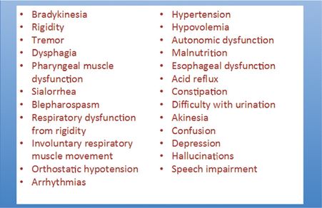
Parkinson’s disease is characterized by the triad of resting tremor, muscle rigidity, and bradykinesia, with autonomic dysfunction. Progressive loss of dopaminergic neurons in the substantia nigra causes a relative deficiency of dopamine in the balance of cholinergic and dopaminergic pathways in the basal ganglia. This results in progressive inhibition of thalamic and brainstem nuclei suppressing cortical motor function. Brainstem nuclei inhibition contributes to posture and gait abnormalities [36–37]. Patients with Parkinson’s disease are more prone to postoperative confusion and hallucinations [38]. See Figure 12.2.

Physiologic changes of Parkinson’s disease.
Preoperative
Preoperative evaluation should include assessment of the patient’s cardiac status and respiratory function. There is an increased incidence of esophageal dysfunction and risk for aspiration in this population. Cognitive dysfunction should be documented and the patient and family informed of an increased risk of POD and POCD. Regional anesthesia can limit postoperative opiate requirements and decrease the risks of POD, POCD, Parkinson’s disease symptoms’ exacerbation, and constipation. Anti-Parkinson medications should be continued in the perioperative period to minimize the development of symptoms. Common medications include:
Carbidopa/levodopa (Sinemet®): converted to dopamine via DOPA decarboxylase, this must be taken enterally on a strict schedule given its one- to three-hour half-life.
Selegeline: type B monoamine oxidase inhibitor (MAO-I). It can cause a significant pressor effect in response to indirect- and direct-acting sympathomimetics. Abnormal glucose metabolism may occur.
Dopamine agonists (ropinrole, priampexole, rotigotine): these can be associated with orthostatic hypotension. Anti-dopaminergic drugs can reverse the action of these medications and should be avoided.
The use of deep brain stimulators (DBS) for treatment of Parkinson’s disease has become common. A generator is most often placed subcutaneously in the chest wall and stimulating leads are tunneled to end in the globus pallidus or subthalamic nucleus. Deep brain stimulation reduces the need for maintenance medication, decreases psychiatric symptoms, and improves motor function and activities of daily living in Parkinson’s disease patients [39]. This prolongs patient survival and decreases nursing home admissions [40]. Magnetic resonance imaging (MRI) is contraindicated after DBS placement as diathermy from MRI has been reported to cause brain death [41]. Monopolar cautery may damage the DBS implant, therefore bipolar cautery should be used if possible. If monopolar cautery must be used, the patient return electrode should be placed away from the generator.
Intraoperative
Intraoperatively, regional techinques are preferable. Direct-acting vasopressors should be available and adequate intravascular volume should be maintained to treat impaired autonomic compensation. Multimodal analgesia with non-narcotic analgesics should be administered, although surgeon input regarding non-steroidal anti-inflammatory drugs use should be sought. Sedation, if needed, can be provided with dexmedetomidine or propofol. If general anesthesia is required, the use of anticholinergic drugs that act centrally should be avoided. A review of commonly used intraoperative drugs and the effects on Parkinsonism is as follows:
Phenothiazines (prochlorperazine), butyrophenones (droperidol), and metoclopramide may exacerbate Parkinsonism. Metoclopramide may temporarily cause drug-induced Parkinson’s disease [42].
Inhalational anesthetics inhibit reuptake of dopamine, affecting spontaneous and depolarization-induced dopamine release. Hypotension is increased due to hypovolemia, norepinephrine depletion, autonomic dysfunction, and arrhythmias [43–44].
Succinylcholine does not seem to induce hyperkalemia in the setting of Parkinson’s disease [45].
Opiates have a variable effect. Opioid-induced muscle rigidity is possibly due to presynaptic inhibition of dopamine release [46]. Morphine was associated with a reduction of dyskinesia at low doses and an increase in akinesia at high doses [47]. Acute dystonia with alfentanil has also been described [48]. Additionally, hydromorphone has been associated with myoclonus in the setting of Parkinsonism [49].
Ketamine is theoretically contraindicated in the setting of Parkinson’s disease, due to an exaggerated sympathetic response.
Propofol may cause dyskinetic episodes or abolition of tremor [50–51]. In a patient having stereotactic ablation or DBS placement, etomidate is the drug of choice.
Postoperative considerations for the patient with Parkinson’s disease
Postoperatively a multimodal analgesic regimen should emphasize non-narcotic agents. Pain control balanced with opiate restriction will limit the risk of postoperative delirium, and pulmonary and cardiac complications. Haloperidol, the traditional medication for control of postoperative delirium, is a strong dopamine antagonist and may worsen Parkinson’s disease symptoms. Therefore atypical antipsychotics would become the first choice for this patient if postoperative delirium should occur. To minimize the risk of POD, patients should have natural light, be kept to a normal sleep/wake cycle, remain normothermic, and be reoriented frequently [29, 34, 52].
The optimal anesthetic for the described patient would be an intraoperative spinal anesthetic with a continuous peripheral nerve block as part of a multimodal analgesic regimen. Benzodiazepines and anticholinergics should be avoided. Propofol and phenylephrine may be utilized for sedation and hemodynamic support intraoperatively. Postoperative opiates should be used after maximizing non-opiate analgesics, dosed orally, and conservatively prescribed.
References
Stay updated, free articles. Join our Telegram channel

Full access? Get Clinical Tree


