Identify the appropriate spinous process. A point 2.5 cm lateral to the superior border of the spinous process is marked.
Lidocaine is infiltrated. Before introducing the Tuohy block needle a 22G finder needle can be used to identify the transverse process (3–4 cm deep). The depth of the finder needle and the direction is noted. A 17 or 18G Tuohy needle is then advanced transverse process (Figure 26.2) to the identified.

A 17 to 18G Tuohy needle is advanced to identify the transverse process.
Once the transverse process is located, the needle is withdrawn to the skin and advanced in slightly caudad/cephalad direction: enough to walk off the transverse process plus 1 cm further than the distance to the depth of the transverse process. A subtle pop of the costotransverse ligament is felt. A loss of resistance syringe can also be used to identify the space, but is not a guarantee of correct placement. Once in the space, a catheter can be inserted 3 to 5 cm into the PVS.
Ultrasound-guided paravertebral block
Paravertebral blocks can also be performed with the use of ultrasound (US) with either a parasagittal or an intercostal approach.
For the parasagittal approach, a low-frequency (3–5 MHz) curved array or high-frequency (5–15 MHz) linear US probe is placed with its long axis parallel to the spinous processes (Figure 26.3). The selected US probe will be dependent on the patient’s body habitus and consequent depth of the PVS.
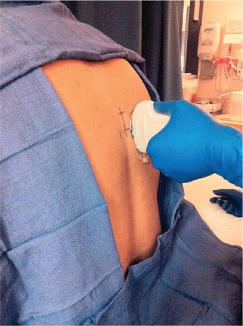
Ultrasound probe orientation and placement.
The probe can be used to scan, starting from the midline, identifying the spinous processes, the lamina, and then the transverse processes. The pleura is identified as the most echogenic, shimmering structure in between the transverse processes (Figure 26.4).

Parasagittal ultrasound approach. Both low-frequency (A) and transverse approach with a high-frequency linear (B) probes may be utilized with this approach.
The costotransverse ligament can also be identified in the same view. A 17 or 18G Tuohy needle is then inserted, in plane to the probe (cephalad–caudad direction) and advanced between the costotransverse ligament and the pleura. Local anesthetic is injected after careful aspiration and the catheter is threaded 4 to 5 cm into the space.
4. Discuss dosing, catheter management, potential complications, and contraindications
Before placing PVBs, the patient’s medical history, clinical course, and thromboprophylaxis regimen should be assessed. A review of the patient’s medications and screening for other injuries (e.g., cervical spine injury) also need to be obtained [15].
After placement of the PVB catheters, certain parameters must be assessed frequently to monitor this patient’s progress and assess the need for continued PVB catheter or dosing adjustments.
Pain scores: numeric pain scores (0–10): For many patients, self-reporting and an attempt to find all sources of pain may be adequate. The following areas should be explored when assessing pain in this population: (1) self-report, (2) a search for all potential causes of pain, (3) observation of patient behavior, (4) surrogate reporting, and (5) an analgesic trial [16].
Vital capacity assessment: Bakhos et al. reported that patients with rib fractures with a vital capacity less than 1.4 L or less than 55% of their predicted vital capacity had a hospital length of stay greater than three days. It was also reported that those with higher vital capacities were more likely to discharge to home at earlier dates. Conversely, those with lower vital capacities were more likely to require discharge to a skilled nursing facility [17].
Incentive spirometry: Inspiratory volume can be determined using the incentive spirometer. A volume less than 15 ml/kg has been considered failure [18].
Cough: Strong or weak [19].
Dosing
Large volumes of local anesthetics should be avoided especially while injecting through the needle in the PVS to prevent complications, should there be epidural spread. Local anesthetics should be administered in aliquots of 5 ml, and preferably only 5 ml, through the needle with the remaining dose being given through the catheter.
Several different dosing schedules have been evaluated. For effective analgesia, relatively dilute concentrations of local anesthetic (0.125% levobupivacaine, infusing at 0.1 ml/kg/h) are associated with adequate analgesia [20]. Lower infusion rates with larger patient-controlled boluses have been compared with higher infusion rates with small patient-controlled boluses and result in similar analgesic outcomes [21].
Contraindications to PVB
Coagulopathy
Infection at the site
Pleurodesis
Allergy to local anesthetic
Complications
Complications associated with paravertebral blocks are hypotension most likely resulting from epidural spread, vascular puncture, pleural puncture, and pneumothorax.
Related posts:


Stay updated, free articles. Join our Telegram channel

Full access? Get Clinical Tree


