Gastrointestinal Endoscopy
Alexander J. Eckardt
Wahid Wassef
Gastrointestinal (GI) endoscopy has evolved into an essential diagnostic and therapeutic tool for the treatment of critically ill patients in the new millennium. The introduction of new endoscopic techniques and rapid improvements in our therapeutic armamentarium has led to major improvements in the management of patients in intensive care units (ICUs) worldwide. This chapter will review general aspects, current indications, contraindications, techniques, complications, and future directions of gastrointestinal endoscopy in the critically ill.
Historical Aspects
The introduction of the Lichtleiter (light conductor) by the German physician Phillip Bozzini [1] in 1804 marks the beginning of the modern era of endoscopy. This endoscope was the first to combine a light source with an optical system and a series of viewing tubes. The pioneering work of Rudolf Schindler led to the introduction of the semiflexible gastroscope. In 1941, Schindler founded the American Gastroscopic Club, now better known as the American Society of Gastrointestinal Endoscopy (ASGE) [2]. Basil Hirschowitz, from Ann Arbor University in Michigan, is recognized for the development of the first fiberoptic endoscope in the late 1950s [3]. Almost a decade later, Overholt [4] extended the use of the fiberoptic endoscope to examine the colon. Therapeutic endoscopy entered the field of gastroenterology in the 1970’s. Wolff and Shinya’s [5] introduction of snare-polypectomy marked the beginning of a new era in gastrointestinal endoscopy, namely the era of therapeutic endoscopy. Continuing technical advances over the past three decades have made therapeutic endoscopy an invaluable tool for the treatment of the critically ill.
Endoscopes
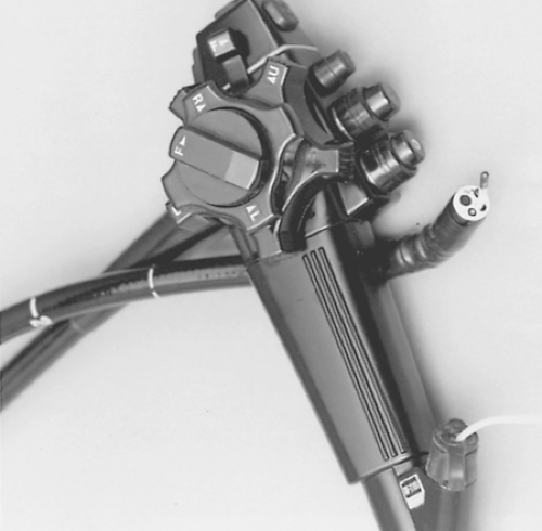
Many of the basic principles of the Lichtleiter still hold true for modern endoscopes. Today’s flexible endoscopes are composed of a control section, an insertion tube, and a connector section [6]. The control section is held in the operator’s left hand. Wheels and buttons on the handle of the instrument control tip deflection (up/down, left/right), suction, and air and water insufflation (Fig. 13-1). The insertion tube is attached to the control section and varies in length, diameter, and stiffness characteristics. It contains one or two instrument channels, air-and water channels, and either a fiberoptic bundle or an electronic video system. The connector section has a light guide, an air pipe, and contacts for the processor/light source.
Standard front-viewing endoscopes used for diagnostic and therapeutic endoscopy are the esophagogastroduodenoscope (EGD), the colonoscope, and the push-enteroscope. They differ in diameter, length, and stiffness. Standard endoscopes are capable of visualizing the upper GI tract to the third or fourth portion of the duodenum, the colon, and terminal ileum. Enteroscopes can visualize parts of the jejunum. The side-viewing duodenoscope is predominantly used for endoscopic retrograde cholangio-pancreatography (ERCP), but can also be useful in the management of other duodenal lesions. ERCP uses fluoroscopy for imaging of the biliary tree. Contrast dye is injected through a catheter that is advanced through the operating channel into the bile duct or pancreatic duct. A sphinctertome is generally used to cut the sphincter of Oddi prior to stone extraction or other interventions (e.g., stenting, dilatation, etc.).
Indications
The indications for gastrointestinal endoscopy in the ICU are summarized in Table 13-1. Generally, endoscopy should only be performed if the results alter patient management. As outlined in Table 13-1, common indications for upper GI endoscopy in the ICU are upper GI bleeding, caustic or foreign body ingestion, and placement of feeding tubes. GI endoscopy in patients with clinically insignificant bleeding or chronic GI complaints should be postponed until their medical/surgical illnesses improve. Endoscopy would only be indicated in critically ill patients with occult blood loss, if anticoagulation or thrombolytic therapy is contemplated.
Upper Gastrointestinal Endoscopy
Upper GI Bleeding
With an estimated 300,000 admissions annually, acute upper GI bleeding (UGIB) is one of the most common medical emergencies [7]. It is defined as the presence of melena, hematemesis, or blood in the nasogastric (NG) aspirate. Studies have shown improved outcomes of UGIBs in critically ill patients with hemodynamic instability or continuing transfusion requirements, if they are managed endoscopically [8, 9]. The distinction between nonvariceal (peptic ulcer, esophagitis, Mallory-Weiss-tear, angiodysplasia, etc.) and variceal lesions (esophageal or gastric varices) is made endoscopically, allowing for targeted therapy [10, 11]. Furthermore, endoscopic stigmata help to prognosticate risk of rebleeding in peptic ulcers, occasionally revealing lesions that require surgical or radiologic intervention [12].
Foreign Body Ingestion
Foreign body ingestions (FBI) occur commonly. They can be divided into two groups: food impactions and caustic ingestion. Food impactions constitute the majority of FBI. Although most will pass spontaneously, EGD should always be performed to look for the underlying cause of obstruction (strictures, rings, etc.). Endoscopic removal of the food bolus is necessary in 10% to 20% of cases, and 1% of patients will ultimately require surgery [13]. Although caustic ingestions only constitute a small number of FBI, they are frequently life-threatening, especially when they occur intentionally in adults. Endoscopy is safe and should be performed for prognostication and triage [14].
TABLE 13-1. Indications for Gastrointestinal (GI) Endoscopy | |
|---|---|
|
Feeding Tubes
Enteral nutrition improves outcomes in critically ill patients and is preferred over parenteral nutrition in patients with a functional GI tract [15]. Although naso- or oroenteric feeding tubes may be used for short-term enteral nutrition, these tubes are felt to carry a higher risk of aspiration, displacement, and sinus infections than endoscopically placed percutaneous tubes. Percutaneous endoscopic gastrostomy (PEG), jejunal extension through a PEG (PEG-J), or direct endoscopic jejunostomy (D-PEJ) [16] are appropriate for many patients in the ICU. They are especially useful in patients with a reversible disease process, likely to require more than 4 weeks of artificial nutrition (e.g., neurologic injury, tracheostomy, neoplasms of the upper aerodigestive tract, etc.) [17]. PEG-J and D-PEJ tubes are appropriate for patients with severe gastroesophageal reflux, gastroparesis, or repeated feeding-related aspiration. Endoscopic gastrostomies or jejunostomies may also be indicated for decompression in patients with GI obstruction [18]. Although this is a technically simple procedure that can be performed at the bedside under conscious sedation, its risks and benefits should always be weighed carefully [19].
Endoscopic Retrograde Cholangio-Pancreatography
The most common indication for urgent ERCP is complicated biliary obstruction from gallstones. Four randomized-controlled trials have studied the role of ERCP in acute gallstone pancreatitis, as summarized in a recent metaanalysis [20]. These and other studies showed that ERCP improves outcomes in the presence of cholangitis, progressive jaundice, or severe pancreatitis [21 22 and 23]. ERCP with sphincterotomy or stenting is also indicated for the diagnosis and treatment of postoperative (hepatobiliary surgery) or trauma-related bile leaks [24, 25, 26 and 27].
Lower Gastrointestinal Endoscopy
Colonoscopy and flexible sigmoidoscopy are used much less frequently than upper endoscopy in the ICU. The main indications for urgent colonoscopy are lower GI bleeding, colonic pseudo-obstruction (Oglivie’s syndrome), volvulus, and evaluation for infection (Cytomegalovirus [CMV], clostridium difficile) in the immuno-compromised, or graft versus host disease (GVHD) in the transplant patient [28, 29].
Lower GI Bleeding
Acute lower GI bleeding (LGIB) is defined as bleeding from a source distal to the ligament of Treitz for less than 3 days and is predominantly a disease of the elderly [30]. Common causes are diverticular bleeding, ischemic colitis, and vascular abnormalities. However, as many as 11% of patients initially suspected to have a LGIB are ultimately found to have an UGIB [31]. Therefore, UGIB sources should always be considered first in patients with LGIB. Once an upper GI source has been excluded, colonoscopy should be performed. Although urgent colonoscopy within 24 to 48 hours has shown to decrease the length of hospital stay [32, 33] and endoscopic intervention is
often successful, 80% to 85% of LGIBs stop spontaneously [34]. If the bleeding is severe or a source cannot be identified at colonoscopy, a technetium (TC) 99m red blood cell scan with or without angiography should be considered [35, 36].
often successful, 80% to 85% of LGIBs stop spontaneously [34]. If the bleeding is severe or a source cannot be identified at colonoscopy, a technetium (TC) 99m red blood cell scan with or without angiography should be considered [35, 36].
Volvulus and Acute Colonic Pseudoobstruction
Acute colonic obstruction is considered a medical emergency. Imaging studies (abdominal plain films, computed tomography [CT] scan) are needed to differentiate “true” mechanical obstruction from acute colonic pseudoobstruction, because these entities are managed differently.
Volvulus
Colonic volvulus is the third most common cause of mechanical colonic obstruction in the western world, following neoplasms and diverticular disease [37]. It presents a “closed-loop obstruction” with risk for ischemia, perforation, and death. Decompressive endoscopy with minimal inflation of air resolves the acute obstruction in the majority of cases (81%) [38]. Despite a high recurrence rate (23% to 57%), colonoscopy is often considered the initial procedure of choice in the absence of intestinal ischemia [38, 39].
Stay updated, free articles. Join our Telegram channel

Full access? Get Clinical Tree


