 Unstable patients with gastroesophageal varices receiving maximal medical therapy
 Endoscopy is unavailable or unsuccessful
CONTRAINDICATIONS
 Esophageal strictures or recent gastroesophageal surgery
 Relative:
 No active bleeding
 Incomplete equipment
 Source of bleeding likely gastric
 General Basic Steps
 Gather supplies
 Prepare patient—intubate
 Placement and gastric balloon inflation
 Traction
 Esophageal balloon inflation
SUPPLIES
 Sengstaken–Blakemore (SB) tube (triple lumen tube) or Minnesota tube (quadruple lumen tube). The fourth port is for suctioning the proximal esophagus.
 Salem Sump (double-lumen nasogastric tube) and silk ties to create necessary fourth lumen (not needed if using a Minnesota tube)
 60-mL Luer lock syringe
 60-mL Piston syringe
 2 Christmas tree catheter adapters
 2 Three-way stopcocks
 2 Heplock caps
 Surgilube
 1 Sterile gauze bandage roll (Kerlix)
 1 L NS (normal saline)
 Kelly clamps (padded)
 2 wall-suction units
 Straight connector
 Manual sphygmomanometer
TECHNIQUE
 Preparation
 Secure the airway. The patient will be intubated in almost all scenarios. Raise the head of the bed to 45 degrees.
 Assemble attachments to gastric balloon and esophageal balloon ports
 Test for air leaks using 60-cc Luer lock syringe
 Gastric balloon—Inflate 250-cc air
 Esophageal balloon—Inflate 60-cc air
 Deflate the balloons completely
 If using an SB tube, create fourth lumen:
 Place the distal tip of Salem Sump 2 cm proximal to the esophageal balloon and secure with silk ties (FIGURE 29.1)
 Placement and Gastric Balloon Inflation
 Lubricate the gastroesophageal balloon tamponade (GEBT). Insert orogastrically so that the 50-cm mark aligns with the patient’s lip. Can insert nasally; however, the oral route is preferred (FIGURE 29.2).
 Confirm placement via air insufflation through gastric port and auscultation for gastric sounds
 Connect gastric port to 60 to 120 mm Hg intermittent suction. Inflate the gastric balloon with 50 cc of air.
 Confirm with chest x-ray that the inflated balloon is in the stomach
 Inflate additional 200 cc of air into gastric balloon, for a total of 250 cc of air
 Affix padded Kelly clamp to gastric balloon port
 Traction
 The proximal end of the GEBT needs to be secured with traction
 Attach Kerlix distal to SB tube ports by creating a slip knot. Secure the opposing end to 1-L NS bag (or similar weight).
 Hang Kerlix over the IV pole, allowing the 1-L NS bag to hang freely, applying traction to the SB tube
 Esophageal Balloon Inflation
 Connect sphygmomanometer to the three-way stopcock on esophageal balloon port
 Inflate the esophageal balloon to 30 to 45 mm Hg (typically 50–70 cc air), using lowest pressure necessary
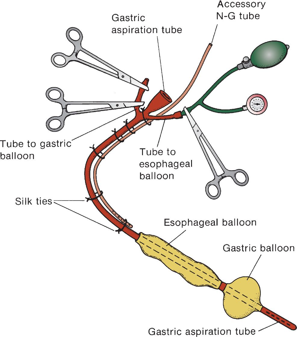
FIGURE 29.1 Modified Sengstaken–Blakemore tube. Also available is the Minnesota tube, which has a built-in esophageal port. (Reused with permission from Yamada T. Textbook of Gastroenterology. 4th ed. Vol 1. Philadelphia, PA: Lippincott Williams & Wilkins; 2003:707.)
Related posts:



Stay updated, free articles. Join our Telegram channel

Full access? Get Clinical Tree


