Melissa A. Hall
Fungal Infections (Superficial)
Superficial fungal infections are common problems. Greater exposure to fungal pathogens is occurring in the health and fitness–minded population, in debilitated patients using systemic antibiotics, in diabetics, and in other patients who are immunocompromised. These fungal infections can cause a primary or secondary infection of the skin that complicates accurate diagnosis of the precipitating condition. Fungal infections are caused typically by dermatophytes or yeast organisms.
Dermatophyte Infections
Definition and Epidemiology
Dermatophytes, the most common fungi that invade the skin and nails, proliferate within the nonviable keratinized tissues—the stratum corneum of the skin, hair, and nails. Three of the most common pathologic dermatophytes are Trichophyton, Microsporum, and Epidermophyton organisms. The infections produced by these dermatophytes are known as tinea, dermatophytosis, or ringworm. The term tinea is derived from the Latin word for worm and was probably chosen because of the common presence of a migrating, circular pattern with the infection. The tinea infection is named by the part of the body that is affected by the dermatophyte.1
Pathophysiology
Fungal infections are usually acquired through inhalation of endemic fungi in the environment, with soil being the natural reservoir.1 They are also transmitted through close contact with an infected person or animal. Tinea corporis is the second most common infection passed from dogs and cats to humans.2 Indirect contact with fomites (infected towels, hats, upholstery, and hairbrushes) may also cause dermatophyte infections. Infections of scalp hair and body surfaces are most frequent during childhood; hand, foot, or nail infections are much more common after puberty.3
Clinical Presentation and Physical Examination
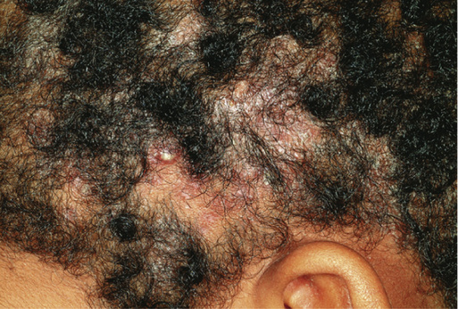
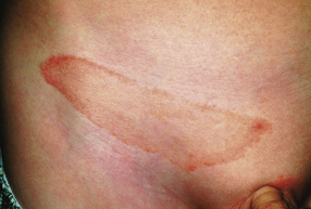
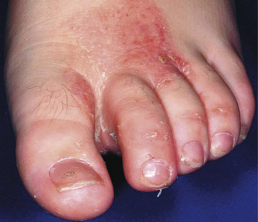
Dermatophyte infections are characterized and named according to their location. Tinea capitis (head or scalp) can be seen initially as patchy, scaly, nonscarring areas of hair loss (Fig. 54-1). Depending on the infectious organism, the lesions may become inflamed, boggy, and pustular. Tinea corporis (body) appears on skin as erythematous plaques and papules in an annular or arciform pattern. Lesions often have slightly elevated borders with central clearing (Fig. 54-2). Tinea cruris (jock itch) appears on the groin and upper inner thigh and extends to the gluteal folds as erythematous scaling patches with raised borders. The scrotum is often spared. Tinea pedis (athlete’s foot) can occur as interdigital scaling, maceration, and fissuring (Fig. 54-3). It can also appear as a mild erythematous scaling eruption that involves the sole and sides of the foot (moccasin distribution). Tinea manus (hand) is often a dry, diffuse, scaly eruption of the palms, with sharply marginated plaques on the dorsum of the hands. The feet are often also involved. Tinea unguium (nail), also called onychomycosis, most commonly manifests as the distal subungual type. The infection begins in the distal nail bed and spreads to infect the nail plate, causing the nail to appear thickened and yellowed, with subungual keratinous debris. Onychomycosis appears on lateral nail margins as a yellow discoloration. Increased nail thickness and distortion usually occur over time.4
Diagnostics
The diagnosis of all fungal infections is based on clinical features and simple diagnostic procedures. The potassium hydroxide (KOH) microscopy preparation is a valuable, cost-effective tool that provides rapid confirmation of many types of fungal infections.5 The key to a reliable KOH preparation is properly obtaining an adequate specimen by scraping the active, leading border of a lesion. The provider should use a No. 15 blade to scrape the lesion gently, collecting the scrapings on a glass slide, and then place several drops of a 10% to 20% solution of KOH directly on the collected scale and apply a coverslip. Next, the provider should gently heat the sample and examine it under a microscope, looking for the diagnostic branching appearance typically seen with these organisms, which indicates a positive result. The provider should note that a negative KOH result does not exclude dermatophyte infections.
Fungal cultures can help detect infection in the absence of a positive KOH result. Dermatophytes can take a few weeks to grow in fungal cultures. A Wood lamp can be used to screen for tinea capitis caused by certain fungal species; these species fluoresce in the long-wave ultraviolent light produced by the Wood lamp. However, Trichophyton tonsurans, the most common cause of tinea capitis in the United States, does not fluoresce.5
Differential Diagnosis
See the Differential Diagnosis box.
Management
The treatment of tinea infections consists of removal of the infecting organisms. Acute, exudative lesions are treated with drying agents such as aluminum sulfate (Domeboro) soaks. Topical antifungal solutions and creams reduce superficial scaling and organisms; keratolytic agents remove the thick scales on the hands and feet, allowing topical antifungal agents to penetrate better. Topical applications are available to treat dermatophyte infections (Table 54-1); these medications include terbinafine (Lamisil), naftifine (Naftin), butenafine (Mentax), clotrimazole (Lotrimin), econazole (Ecoza), ketoconazole (Nizoral), luliconazole (Luzu), miconazole OTC, oxiconazole (Oxistat), sertaconazole (Ertaczo), ciclopirox (Penlac), and tolnaftate (Tinactin). The products should be continued 1 week after clearing of the lesions to discourage recurrence; however, recurrence of tinea infections is common.6 Oral ketoconazole (Nizoral) should be avoided because of risks of hepatotoxicity and serious drug interations.7 Multiple randomized controlled trials provide evidence that tinea pedis (athlete’s foot) can be treated successfully with over-the-counter terbinafine.8
TABLE 54-1
Examples of Treatments
| Recommended Application to Affected Areas | Indicated for | |||||
| Tinea (Pedis Cruris, or Corporis) | Candidiasis | Tinea Versicolor | Tinea Unguium | Tinea Capitis | ||
| Clotrimazole (Lotrimin) | Twice daily | X | X | X | ||
| Miconazole (Monistat-Derm) | Once to twice daily | X | X | X | ||
| Ketoconazole (Nizoral) | Once daily | X | X | X | X | |
| Oxiconazole (Oxistat) | Once daily | X | ||||
| Ciclopirox (Loprox) | Twice daily | X | X | X | ||
| Nystatin (Mycostatin) | Twice daily | X | ||||
| Butenafine (Mentax) | Once daily | X | X | |||
| Econazole (Spectazole) | Once to twice daily | X | X | X | ||
| Itraconazole (Sporanox) | Once daily | X | X | X | ||
| Griseofulvin | Once daily | X | X | X | ||
| Luliconazole (Luzu) | Once daily | X | ||||
| Fluconazole (Diflucan) | Weekly | X | X | X | ||
| Terbinafine (Lamisil) | Once to twice daily | X | X | X | X | |
| GENERAL CONSIDERATIONS | ||||||
| Clinical improvement may be seen fairly soon after initiation of treatment. Twice-daily applications, when indicated, should be done morning and evening. In general, all infections should be treated for 2 weeks after infection has resolved to reduce the possibility of recurrence. Tinea pedis, tinea unguium, and tinea capitis require 6 weeks or more of treatment. | ||||||


Stay updated, free articles. Join our Telegram channel

Full access? Get Clinical Tree


