 To remove external foreign bodies (FBs) in the eye
 Symptoms include pain, redness, increased tearing, or FB sensation
CONTRAINDICATIONS
 Caution and care must be taken if globe rupture is suspected especially when the history includes flying particles or high-velocity projectiles
LANDMARKS—FIGURE 85.1
 General Basic Steps
 Preparation
 Inspection
 Stain
Stain
 Slit lamp examination
 Removal of FB
TECHNIQUE
 Patient Preparation
 Place 0.5% tetracaine or 0.5% proparacaine drops in the eye (may use in both eyes to reduce the blink reflex)
 In case of intense blepharospasm, administer an ipsilateral facial block
 Inspection—FIGURE 85.2
 Examine the conjunctiva and cornea carefully. Do not assume there will only be one FB.
 Carefully examine behind both eyelids
 Lower eyelid: Pull down the lower eyelid and ask the patient to look up
 Upper eyelid: Using a cotton-tipped applicator as a fulcrum, carefully pull the eyelashes down and out to evert the lid
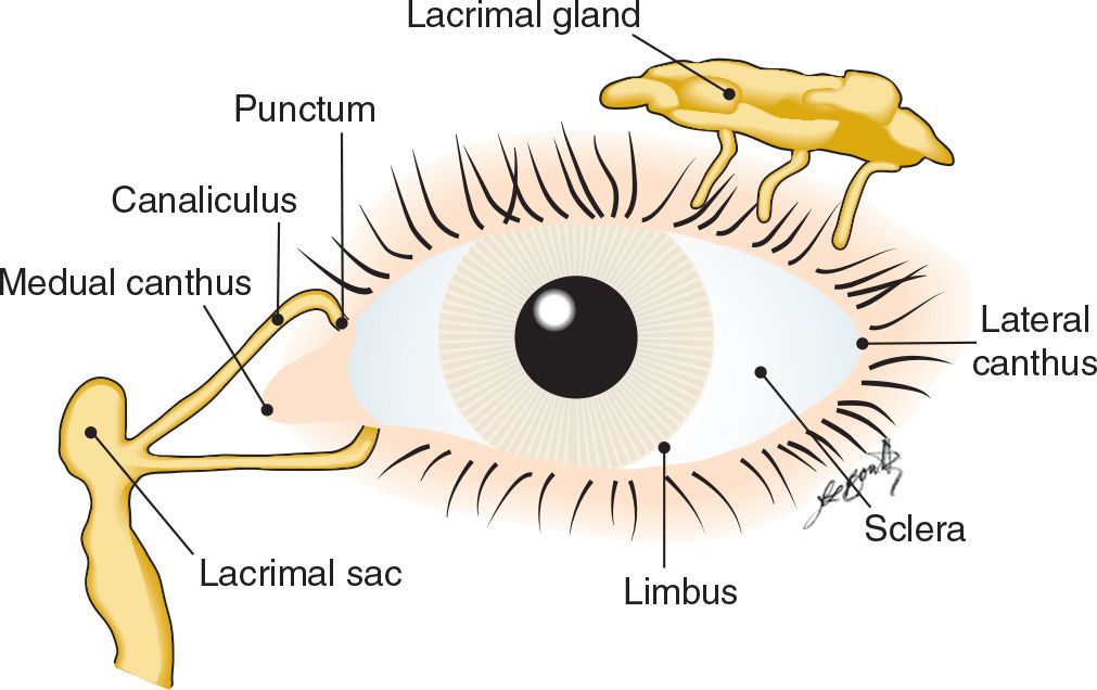
FIGURE 85.1 Periorbital structures. (From Knoop KJ, Dennis W. Eye trauma. In: Wolfson AB, ed. Harwood-Nuss’ Clinical Practice of Emergency Medicine. 6th ed. Philadelphia, PA: Lippincott Williams & Wilkins; 2015:174, with permission.)
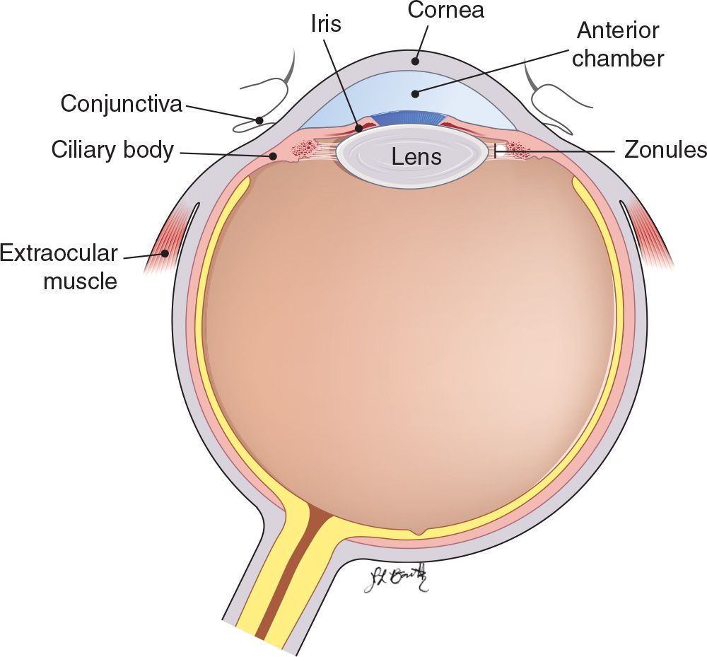
FIGURE 85.2 Cross section of the eye. (From Knoop KJ, Dennis W. Eye trauma. In: Wolfson AB, ed. Harwood-Nuss’ Clinical Practice of Emergency Medicine. 6th ed. Philadelphia, PA: Lippincott Williams & Wilkins; 2015:175, with permission.)
 Fluorescein Stain
 Gently touch the fluorescein strip to the lower eyelid conjunctiva and ask the patient to blink two to three times. Wipe away the excess.
 Inspect the cornea for abrasions (fluorescein when taken up by the alkaline Bowman membrane, will fluoresce with a cobalt-blue light). Patients with corneal abrasions may have FB sensation in the absence of a retained FB.
 Positive Fluorescein Stain (FIGURE 85.3)
 Slit Lamp Inspection
 Examine the fluorescein-stained cornea under the blue light of a slit lamp
 Vertical linear lesions on the cornea should raise suspicion for an FB under the eyelids
 If you see an FB extending through the full thickness of the cornea, consult an ophthalmologist
 Signs of an intraocular FB may be subtle or absent. Look carefully for the following:
 Irregular pupil
 Shallow anterior chamber
 Collapsed iris
 Positive Seidel test (extrusion of fluorescent material from the cornea)
 Hyphema
 Lens opacification
 Decreased intraocular pressure
FOREIGN BODY VISIBLE
 Swab
Swab
 If easily visualized, remove the particle with a moist sterile cotton-tipped applicator or nasopharyngeal swab
 Irrigation
 FB may be flushed out when the eye is irrigated gently with a stream from an Angiocath connected to a syringe containing saline
 If more copious irrigation is required, consider commercial devices such as the Morgan lens
 Embedded FB
 Cornea can be gently scraped with a small 25- or 27-gauge needle, attached to a syringe for stability (under the slit lamp)
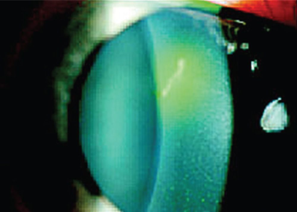
FIGURE 85.3 Corneal abrasion stained with fluorescein under cobalt-blue light. (From Wilson SA, Last A. Management of corneal abrasions. Am Fam Physician. 2004;70(1):123–128. http://www.aafp.org/afp/2004/0701/p123.html. Accessed March 30, 2014.)
Related posts:



Stay updated, free articles. Join our Telegram channel

Full access? Get Clinical Tree


