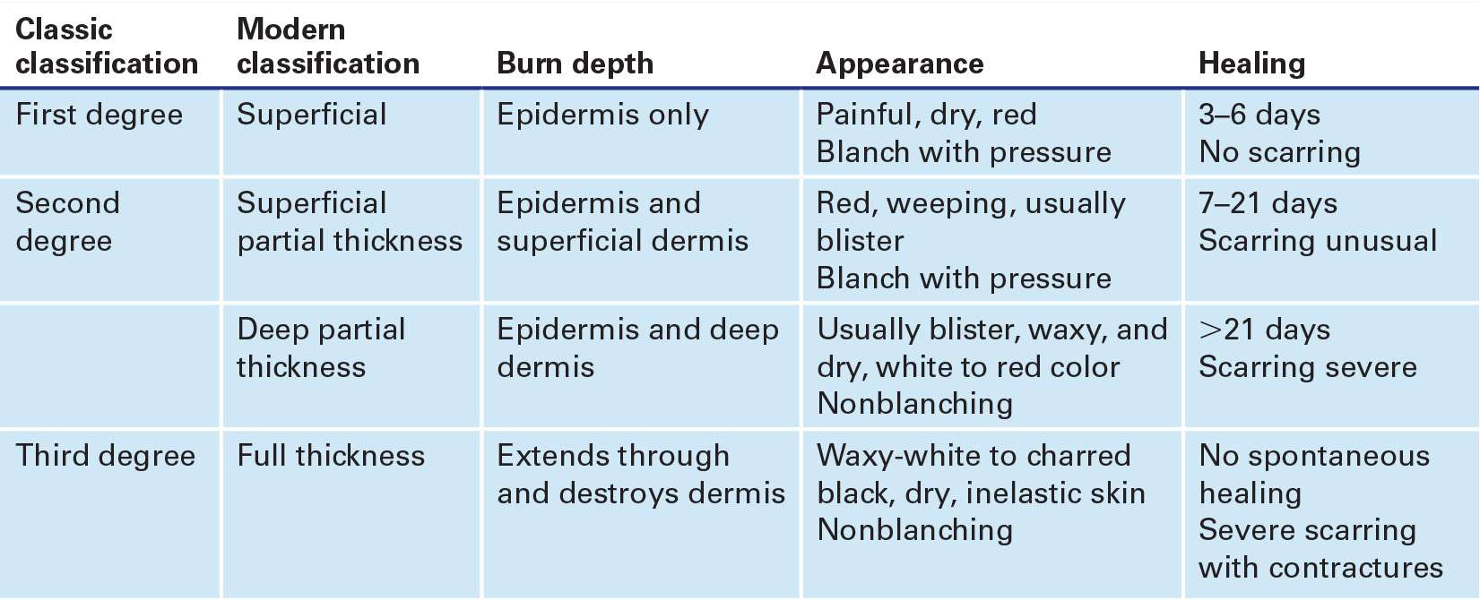
 Used to decompress accumulated edema under tight, unyielding eschar following full-thickness burn (classic and modern classifications of burns are given in TABLE 74.1)
 Circumferential extremity burn with evidence of neurovascular compromise:
 Cyanosis
 Deep tissue pain
 Progressive paresthesia
 Decreased or absent pulses
 Elevated compartment pressure
 Decreased arterial flow on Doppler ultrasonography
 Pulse oximetry <95% of affected extremity (without systemic hypoxia)
 Thoracic burn with evidence of respiratory compromise due to eschar
 Circumferential neck burn
 Abdominal burn with evidence of increased intra-abdominal pressure (usually estimated by bladder pressure)
 Circumferential penile burn
CONTRAINDICATIONS
 No evidence of tissue hypoperfusion on physical examination
 Normal findings on arterial Doppler ultrasonography
 Adequate respiration despite eschar
 No evidence of increased intra-abdominal pressure
RISK/CONSENT ISSUES
 Often difficult to obtain consent from major burn victims; escharotomy is a life-saving procedure and should be performed even if informed consent from the patient cannot be obtained
 Procedure can cause pain (local and systemic analgesia will be provided)
 Risk of bleeding (minimized with proper technique)
 Whenever the skin is broken, there is potential for introducing infection (sterile technique will be utilized)
BURN DEPTH CLASSIFICATION |
LANDMARKS
Escharotomy sites are depicted in FIGURE 74.1.
TECHNIQUE
 General Basic Steps
 Airway, breathing, and circulation (ABC)
 Consider early intubation
 Fluid resuscitation
 Analgesia
 Tetanus prophylaxis
 Wound care
 Escharotomy
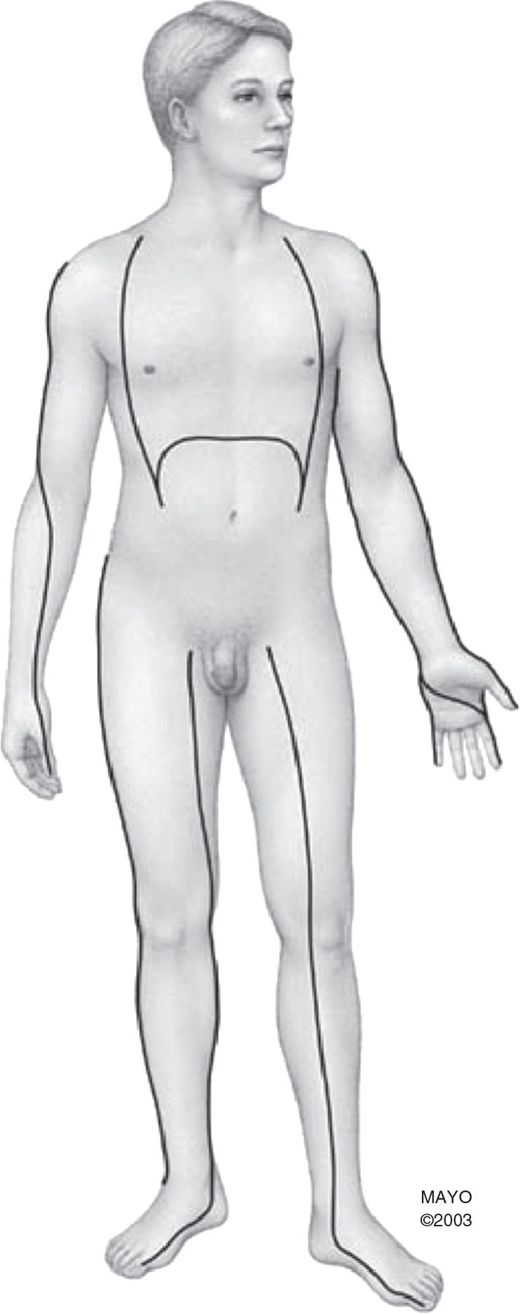
FIGURE 74.1 Escharotomy sites. (From Haro LH, Miller S, Decker WW. Burns. In: Wolfson AB, ed. Harwood-Nuss’ Clinical Practice of Emergency Medicine. 6th ed. Philadelphia, PA: Lippincott Williams & Wilkins; 2014:315, with permission.)
BURN MANAGEMENT
 First ensure ABC and administer supplemental oxygen
 Strongly consider endotracheal intubation if:
 Burns to the face and neck are present
 Soot in and around the mouth and nose
 Hoarseness, stridor, wheezing, or development of acute coughing
 Carbonaceous sputum
 Give intravenous fluids for resuscitation (for moderate to major burns)
 Use Parkland formula: Ringer lactate 4 mL × weight (kg) × % of total body surface area (TBSA) burned (excluding superficial burns)
 Give ½ of total volume over the first 8 hours from time of burn injury
 Give second ½ of total volume over the following 16 hours
 Titrate to maintain blood pressure and urine output of at least 1 mL/kg/hour
 Continue maintenance fluids in addition
 Place urinary catheter to monitor adequate resuscitation (FIGURE 74.2)
 Provide pain management with frequent pain assessment
 Acetaminophen and nonsteroidal anti-inflammatory drugs (NSAIDS) with or without opioids for superficial burns
 Opioids are necessary for partial-to-full thickness burns
 Administer tetanus prophylaxis
 Wound care, if not delaying transfer to burn unit:
 Use sterile technique
 Clean with mild soap and tap water
 Debride sloughed or necrotic skin; avoid extensive debridement
 Remove ruptured blisters
 Intact blister management is controversial; it is recommended to unroof cloudy blisters or those where rupture is imminent (e.g., over joints)
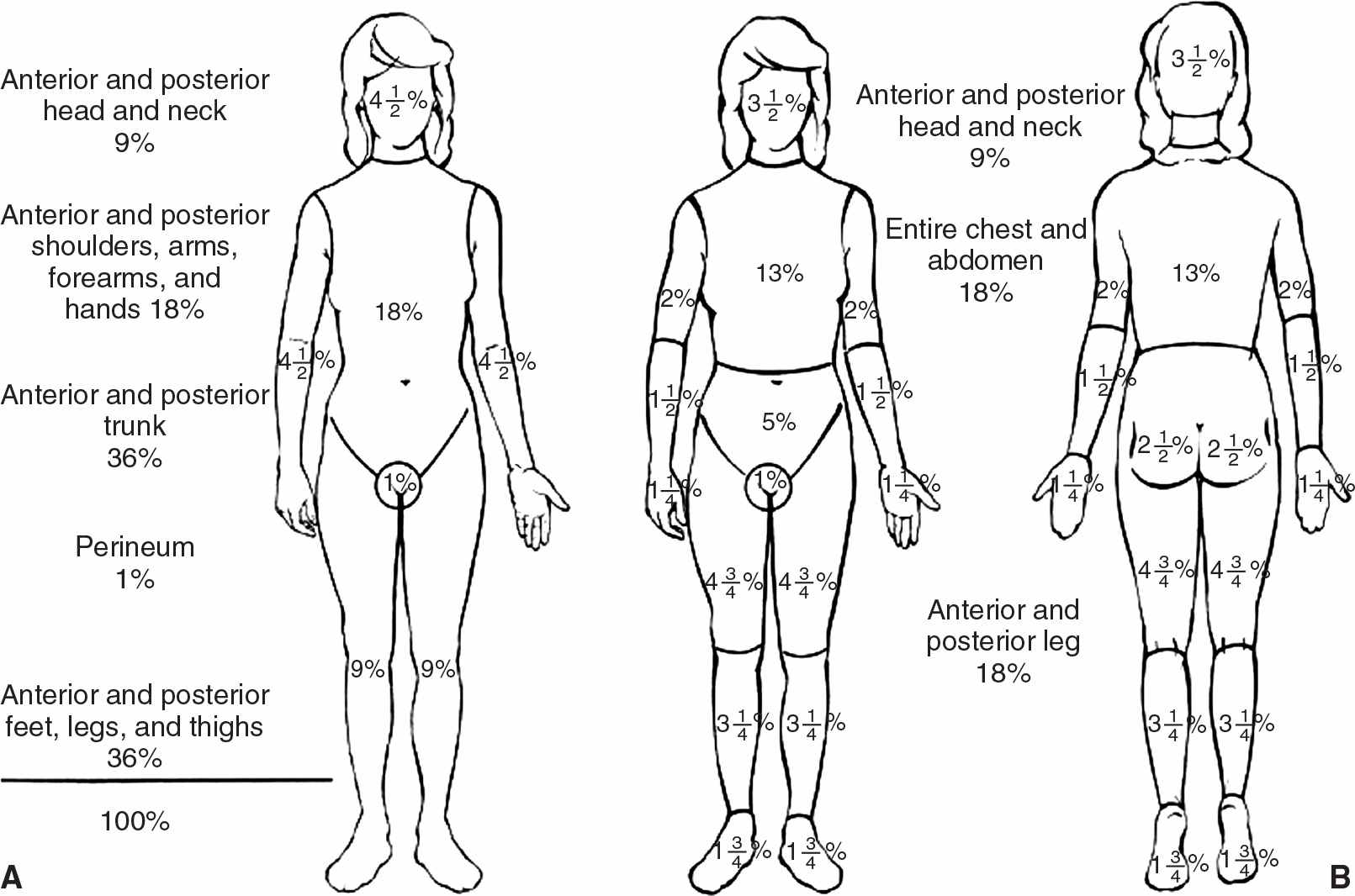
FIGURE 74.2 Methods to evaluate percentage of body surface area burned. A: Rule of Nines. B: Lund and Browder chart. (From Haro LH, Miller S, Decker WW. Burns. In: Wolfson AB, ed. Harwood-Nuss’ Clinical Practice of Emergency Medicine. 6th ed. Philadelphia, PA: Lippincott Williams & Wilkins; 2014:1103, with permission.)
Related posts:



Stay updated, free articles. Join our Telegram channel

Full access? Get Clinical Tree


