CHAPTER 5
Equipment Used in Pain Management
INTRODUCTION
Interventional pain management involves the use of image-guided procedures as well as targeted delivery of medications to modulate pain, symptoms, and disease. To facilitate these procedures, an interventionalist has to utilize a variety of equipment, ranging from simple needles for a trigger point injections to the use of an MRI or a CT scanner for more complex procedures.
Without proper equipment, the procedures cannot be done in a reliable manner and are basically described as “blind” techniques. This chapter covers various types of needles and specialized catheters and syringes. Some major equipment such as a fluoroscopy machine is covered elsewhere in this atlas (see Chapters 1 and 4).
RELEVANCE OF ANATOMY
Although it is necessary to consider the anatomical variations in males, females, adults, and children, the basic need for most of the equipment remains the same. However, depending on the patient’s size and type of a procedure, one must consider the proper type and length of needles, anticipate difficulties, and be prepared for possible complications in case ideal equipment is not available. For example, a larger patient would need a longer needle (and choosing a shorter needle may lead to multiple or failed attempts as well as improper placement of the needle), a sharp needle instead of a pencil point needle may lead to nerve damage, a spinal needle used for epidural access would lead to higher rates of dural punctures. Without proper equipment for guidance of a needle (such as a fluoroscopy unit or an ultrasound unit), the needle may end up in an unintended location and even cause unintentional trauma to tissue.
BASIC CONCERNS AND CONTRAINDICATIONS
Based on the procedure to be performed, it is inappropriate to use certain equipment such as a long needle for a shallow procedure which can lead to higher rate of complications.
TYPES OF EQUIPMENT
Needles (Nonepidural Placements)
Quincke (Figure 5-1)
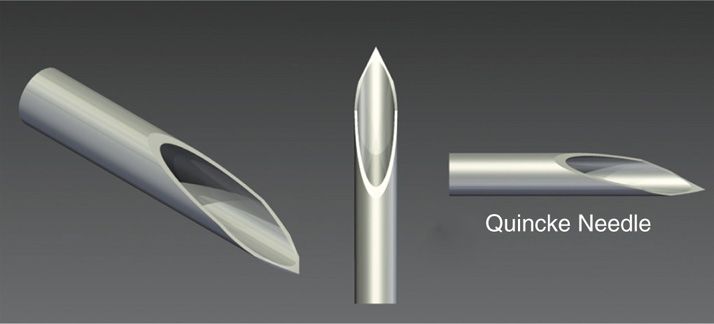
Figure 5-1. Quincke needle—note the sharp tip of the needle which has a cutting edge. (Used with permission from Aakash Patel.)
• Most common type of bevel seen in needles.
• It is a sharp bevel that facilitates penetration of the skin.
• Various needles that have such bevels are intended for intramuscular and intravenous access, biopsies, subcutaneous drug delivery, etc.
• Spinal needles are mainly available with this type of bevel and are commonly used for several interventional procedures.
Pencil point (Figure 5-2)
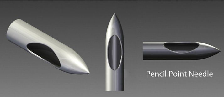
Figure 5-2. Pencil point needle. (Used with permission from Aakash Patel.)
• This needle has a tip that is conical in shape.
• The bevel is on the side of the needle shaft at the conical tip.
• Although considered a “blunt” type needle, it can easily penetrate delicate structures such as a vein or even an artery if the vessel is fixed and immovable.
Bullet tip (Figure 5-3)
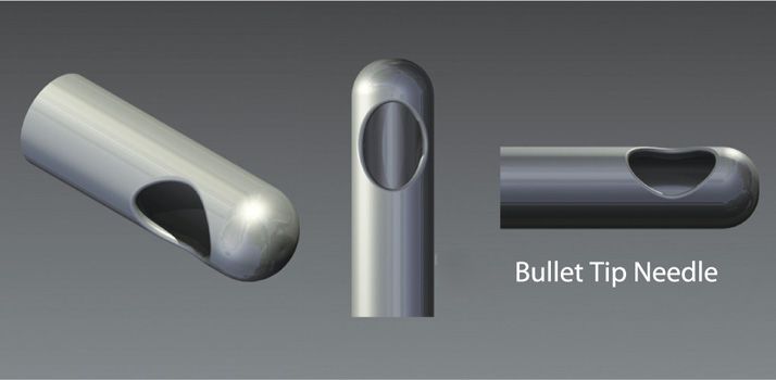
Figure 5-3. Bullet tip needle. (Used with permission from Aakash Patel.)
• Similar to the pencil point, but is completely blunt at the tip.
• Safer for certain procedures that are in the close proximity of nerves and blood vessels.
• Bevel is at the side of the tip.
• A sharp-tipped introducer is a must as this tip cannot penetrate the skin.
“B” bevel
• Commonly used for nerve blocks. The relatively short and noncutting tip makes it a safer choice for a peripheral nerve block.
Chiba needle (Figure 5-4)
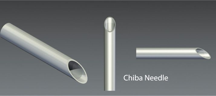
Figure 5-4. Chiba needle—note the angled bevel with a noncutting edge. (Used with permission from Aakash Patel.)
• It has a very short bevel with a noncutting tip.
• Suitable for deeper procedures when a needle has to traverse delicate issues on its way to the target such as a lumbar sympathetic block or celiac plexus block.
Day needle (Figure 5-5)
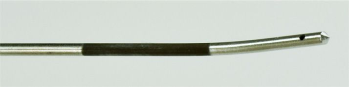
Figure 5-5. Day needle. (Used with permission from Epimed International, Inc.)
• Bullet tip Coudé needle (see below) used for various interventional procedures in the vicinity of delicate tissues.
• The bullet tip minimizes the damage to such tissues as it has a noncutting tip.
Huber needle (Figure 5-6)
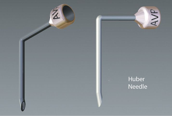
Figure 5-6. Huber needle—the tip has a noncoring edge, which is safer for the silastic injection port. (Used with permission from Aakash Patel.)
• Used for accessing a sub-cutaneous reservoir or a port during a continuous infusion such as used in a long term epidural infusion.
• Angled shaft helps keep the needle flat against the skin while it is placed within the port.
• The tip (similar to a Touhy needle) is also specially designed to be “noncoring” so that it does not damage the silastic material over the entry port.
Needles (Epidural Placements)
Touhy needle (Figure 5-7)
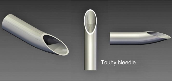
Figure 5-7. Touhy needle—the slight curve helps guide a catheter at an angle and provides a slightly blunter edge to prevent a dural puncture. (Used with permission from Aakash Patel.)
• A blunter tip as the opening is slightly on the side and the tip is curved.
Related posts:


Stay updated, free articles. Join our Telegram channel

Full access? Get Clinical Tree


