Figure 5.1
An intraoral view of erythematous tonsils with debris in the swollen crypts. Follicular tonsillitis
Key Features of History and Examination
Sore throat, pain, and difficulty on swallowing
Past history of sore throats/tonsillitis
Referred otalgia
Fever and malaise
Bad breath
On examination – enlarged congested tonsils ± white exudates on tonsils; palpable and sometimes tender cervical lymphadenopathy
In the early stages, glandular fever is often hard to distinguish from acute bacterial tonsillitis.
Principles of Acute Management
Analgesia, antibiotics, and maintain hydration.
Oral or IV antibiotics depending on patient’s general state.
Penicillin V/clarithromycin are the preferred oral antibiotics. Amoxicillin is best avoided in suspected glandular fever because it can cause severe skin rash.
Often patients have difficulty in swallowing. Anesthetic gargles temporarily relieve the odynophagia. Soluble oral or rectally administered analgesia is preferable.
Patients who are unable to swallow fluids adequately will require hospital admission for IV antibiotics, analgesics, and IV hydration.
Investigations: FBC, U&E, glandular fever screening test, LFTs (if glandular fever suspected) and throat swab.
In patients with glandular fever one or two doses IV steroid injections (dexamethasone 8 mg) can give significant symptomatic relief.
General Discussion
Acute tonsillitis is a common ENT problem seen in emergency department and general practice. Tonsillitis can affect any age group but is most common in young children and teenagers. Common organisms causing acute bacterial tonsillitis are Streptococcus species, H. influenza, and S. aureus.
Viral tonsillitis is also common but glandular fever (caused by EB virus) deserves special attention owing to the severity of the symptoms and the prolonged course of infection and the sequel.
Glandular fever (infectious mononucleosis) is caused by Epstein-Barr virus. It is common in teenagers and young adults. It is usually a self-limiting condition, but the symptoms of tonsillitis are more severe and prolonged. Marked cervical lymphadenopathy (hence the name glandular fever) and occasional hepatosplenomegaly occur in this condition. Diagnosis is confirmed by Monospot or Paul-Bunnell blood test.
Complications of Acute Tonsillitis
Quincy (Peritonsillar Abscess)
Quincy denotes formation of an abscess in the peritonsillar tissue in the soft palate, displacing the uvula to the opposite side. It is almost always unilateral. Patient presents with trismus and drooling of saliva due to inability to swallow.
Aspiration of abscess with a wide bore needle or incision and drainage along with analgesics and IV antibiotics is the mainstay of the treatment. Local anesthetic spray is not generally required. The point where an imaginary line along free edge of anterior tonsillar pillar joins another imaginary line along base of uvula would be the ideal site for aspiration/I&D. Further aspirations may be required in the following 24–48 h as abscess is likely to recur.
Parapharyngeal Abscess
It can occur as a rare complication of acute tonsillitis. It is more common in immunocompromised patients and in patients with indiscriminate use of steroids for acute tonsillitis (often symptoms are masked). It can be a result of spread of infection into parapharyngeal space or a result of lymph node suppuration. It usually presents with diffuse swelling in the neck or in the lateral pharyngeal wall. Early assessment of airway is important. Patients will need ultrasound examination of neck to locate the site and size of abscess. Treatment includes broad-spectrum IV antibiotics with anaerobic cover and drainage of the abscess (usually external approach required).
Acute Airway Obstruction
Rare complication can occur as a result of significant enlargement of tonsils/adenoids in glandular fever. Patient may develop severe sleep apnea. IV steroids can resolve the airway obstruction dramatically.
Miscellaneous Throat Infections
Acute Pharyngitis
This is a common and self-limiting problem. Patient presents with sore throat and irritation. Dysphagia is rare. Voice is unaffected. On examination, congested/inflamed pharyngeal mucosa is identified.
Principles of Management
Need simple remedies – analgesia and rehydration. Saline/anesthetic gargles give symptomatic relief.
Rarely antibiotics are needed. Admission not required unless dysphagia/odynophagia significant.
Acute Laryngitis
This is a common and self-limiting problem. Inflammation of larynx can occur in isolation or in conjunction with URTI. Noninfective causes include voice abuse and acid reflux.
Patient presents with sore throat, pain on speaking or swallowing, hoarseness of voice/loss of voice, and occasionally cough.
Principles of Management
Reassurance, voice rest, analgesics, and cough suppressants. Avoid gargling or whispering as both can aggravate laryngitis.
Ludwig’s Angina
This is an uncommon but potentially life threatening infection of the floor of mouth, which usually results from a dental infection. It is more common in adults. It can cause acute airway problem by upward and backward displacement of tongue due to swelling/abscess in floor of mouth tissues.
Patient presents with trismus, drooling, dysphagia, and high fever. Bimanual palpation of floor of mouth confirms firm thickening of tissues.
Principles of Management
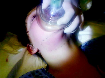
Treat the infection with high dose IV broad-spectrum antibiotics and maintain IV hydration. If airway problem is imminent, secure airway with nasopharyngeal intubation or tracheostomy, if necessary (Fig. 5.2).


Figure 5.2
A young male who presented with progressive submandibular swelling from infection causing airways compromise
Stridor
Case Scenario
A 3-year-old child was brought to the emergency department with noisy breathing, drooling saliva, and high fever. The child was unable to lie flat and was noted to be sitting forward and using accessory muscles of respiration.
Key Features of History and Examination
Epiglottitis and Supraglottitis
Acute inflammation of epiglottis and supraglottic structures is a serious condition occurring in children between the ages of 1–5 years, caused by H. influenza type B bacteria. It can lead to airway obstruction very rapidly. Since the routine HIB vaccination, this condition is less commonly seen in children in developed countries. Adults are more commonly affected these days and often present with supraglottitis.
Preceding URTI, high temperature muffled voice and rapid onset of stridor indicate possible epiglottitis. Do not attempt to examine the throat and do not distress the child for IV access, as this may aggravate the stridor and cause complete airway obstruction. When the diagnosis of epiglottitis is certain, X-raying would be a waste of time, as it does not provide further information. Summon the senior anesthetist and ENT surgeon on call. If the child tolerates, give oxygen by mask and nebulized adrenaline (4 ml of 1 in 10,000) to reduce the edema. Patient is moved to the operating theater at the earliest where airway is best secured by endotracheal intubation, and when this fails, emergency tracheostomy may be necessary.
Discussion
Stridor – high-pitched noise produced by obstructed airway.
Inspiratory stridor – obstruction at the level of vocal cords or above (supraglottic obstruction)
Stay updated, free articles. Join our Telegram channel

Full access? Get Clinical Tree


