CHAPTER 21 Elbow blocks
Clinical anatomy
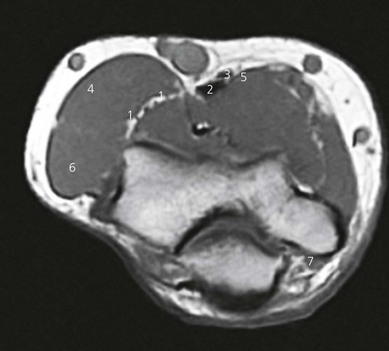
The three major nerves of the forearm can be blocked at the elbow (Fig. 21.1), and also the three cutaneous nerves of the forearm.
The median nerve, which lies anteromedially to the brachial artery near the axilla, crosses medial to the artery in the arm. At the elbow it lies between the tendons of brachialis and pronator teres, and deep to the bicipital aponeurosis. It passes between both heads of pronator teres to enter the forearm (Fig. 21.2). The median nerve provides sensory innervation to the lateral half of the palm, flexor aspect of the thumb, index finger, middle finger, and radial side of the ring finger.
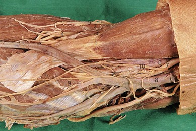
The radial nerve passes between the long and medial head of the triceps to travel around the humerus and emerges on the anterior aspect of the arm above the brachioradialis. It passes deep to the brachioradialis over the lateral epicondyle, where it divides into a deep and a superficial branch (Fig. 21.2). The radial nerve provides sensation for the radial half of the dorsum of the hand, the back of the thumb, and part of the dorsum of the index finger.
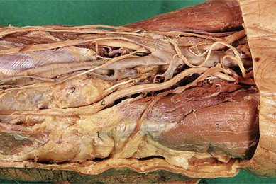
The ulnar nerve leaves the brachial artery halfway down the arm and passes behind the medial epicondyle in a fibrous tunnel. It passes between the two heads of flexor carpi ulnaris to enter the forearm (Fig. 21.3). The ulnar nerve provides sensation for the ulnar half of the dorsum of the hand, little finger, and ulnar side of the ring finger.
Surface anatomy
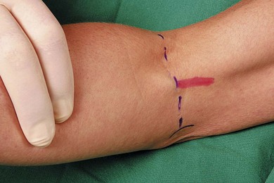
Important structures include the medial and lateral epicondyles of the humerus, olecranon process, intercondylar skin crease, biceps tendon, brachioradialis muscle, and brachial artery (Fig. 21.4). The biceps tendon can be palpated with elbow flexion beneath the intercondylar crease, and it runs laterally to the head of the radius. The brachioradialis muscle can be palpated lateral to the biceps tendon. The brachial artery is palpated medial to the biceps muscle.
Sonoanatomy
The examination begins with the patient supine, the arm abducted, the forearm supinated, and the wrist halfway between pronation and supination (Fig. 21.5). A systematic survey should be performed from superficial to deep and medial to lateral. A high frequency ultrasound transducer is used. With a transverse orientation, the median nerve is found medial to the brachial artery at the elbow as a hyperechoic structure (Fig. 21.6). This is on the medial side of the antecubital fossa. The radial nerve, after winding around the humerus, descends in the intermuscular septum between the brachialis and brachoradialis initially, and then the extensor carpi radialis. The radial nerve is easily seen with ultrasound, as the deep and superficial branches between the brachioradialis and brachialis muscles at the elbow. This nerve appears as a hyperechoic oval structure on the radial side of the antecubital fossa. The two key structures to identify include the hypoechoic brachioradialis and brachialis muscles. The radial nerve lies in the fascial thickening between these (Fig. 21.7). The ulnar nerve is seen posterior to the medial epicondyle at the elbow level in the condylar groove (Fig. 21.8).
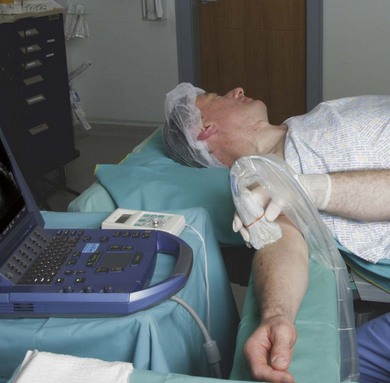
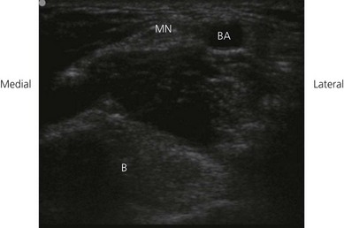
Figure 21.6 Sonosatomy relevant to median nerve block at the elbow. MN: median nerve; BA: brachial artery; B: bone.
Related posts:


Stay updated, free articles. Join our Telegram channel

Full access? Get Clinical Tree


