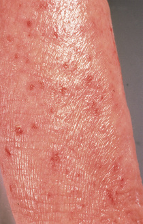
Cené L. Gibson Eczematous dermatitis, or atopic dermatitis (AD), is a pruritic inflammatory skin disorder characterized by exacerbations and remissions of dry, itchy red skin. Onset of the disorder is most common at 3 to 6 months of age, affecting 1 in 10 children and 1 in 10 to 14 adults in the United States.1 AD is also associated with other atopic (immunoglobulin E [IgE]) diseases (e.g., asthma, allergic rhinitis, urticaria, or acute reactions to foods).1–3 AD affects persons of all races, with the international prevalence on the rise.4 Patients with a tendency to develop these conditions are referred to as atopics. Many atopic patients also have a family history of atopy. AD is often called “the itch that rashes.” Patients initially are bothered by incessant itching, scratch an area, and then develop a rash at the site of scratching. Eventually, lichenification may develop at the site if it remains untreated. The primary cause of AD continues to be poorly understood. Primary immune dysfunction resulting in IgE sensitization and/or a primary defect in the epithelial barrier with resultant secondary immunologic dysregulation have been proposed as potential underlying causes of AD.4 AD is characterized by pruritic, erythematous, dry patches of skin, often with scale. Linear excoriations may be seen as a secondary change (Fig. 53-1). The borders of eczematous lesions are not initially well defined. Crusting and oozing are common. Thickened skin with well-defined skin markings (lichenification) may develop in long-standing lesions as the result of scratching. In infants, eruption may involve the cheeks, scalp, forehead, and extensor extremities.1 In adults, eczema or AD may be generalized, with a tendency to develop lesions on the face, neck, flexural folds, wrists, and dorsa of the feet. AD is a clinical diagnosis that is based on a careful history and examination. Seborrheic dermatitis can be differentiated from AD by its presentation and distribution. Seborrheic dermatitis typically manifests as nonpruritic, mildly erythematous plaques with waxy, yellow scale on the face, postauricular area, and scalp. Psoriasis is characterized by well-demarcated, intensely erythematous plaques with characteristic overlying silvery scale, usually located on the scalp, elbows, and knees. Scabies typically is a poorly defined pruritic eruption, often with linear burrows in the web spaces of the fingers. The breasts and genital areas are also often involved. The condition is commonly complicated by eczematous changes from nocturnal scratching and rubbing. The diagnosis is confirmed by scraping of a burrow and microscopic identification of mites, eggs, or feces. Molluscum contagiosum lesions are small, dome-shaped papules with central umbilication. They are not easily confused with AD, but patients with molluscum often develop dermatitis at the site of the lesions. Tinea (or superficial fungal infection) lesions have a sharply demarcated border with scale at the edge and central clearing. They are usually limited in number and sometimes form an arciform array. A scraping of the border of the lesion and treatment of the removed sample with potassium hydroxide (KOH) reveal hyphae on microscopic evaluation. The diagnosis of AD is differentiated on the basis of the presence of xerosis, age at presentation, history of atopic diseases, early onset, and a chronic relapsing course.4 The presentation of allergic contact dermatitis depends on the offending substance. The severity of inflammation associated with contact dermatitis is dependent on the concentration of the irritant and the length of exposure.4 Careful patient history will often reveal potential exposures. Immunodeficiency should be considered in infants with severe itching in the setting of recurrent infection. Mycosis fungoides manifests as hypopigmentation with dermatitis. Diagnostics Atopic Dermatitis
Eczematous Dermatitis (Atopic Dermatitis)
Definition and Epidemiology
Pathophysiology
Clinical Presentation and Physical Examination
Diagnostics and Differential Diagnosis
Eczematous Dermatitis (Atopic Dermatitis)
Chapter 53