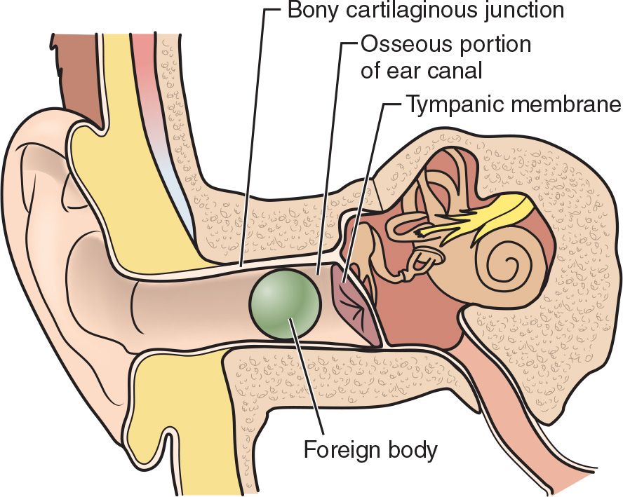
 Removal of foreign matter lodged within the external auditory canal (FIGURE 86.1)
 General Basic Steps
 Preparation
 Irrigation
 Removal
TECHNIQUE
 Patient Preparation
 Place patient in a supine position
 Restrain patient and immobilize the head
 Consider sedation for patients unable to cooperate with the procedure
 Irrigation
 Before the procedure, perform pneumatic otoscopy in patients with suspected tympanic membrane (TM) perforation to document an intact TM
 Pull the pinna superiorly, posteriorly, and laterally in order to straighten the external auditory canal and allow for a more complete visualization of the foreign matter and TM
 Contraindications for irrigation include patients with suspected or known TM or tympanostomy tubes and organic foreign bodies (FBs) because water may cause the object to swell in the external auditory canal
 Attach a 20-mL syringe to a 16- or 18-gauge intravenous catheter or a 2-inch section of butterfly needle tubing (cut off the needle assembly)
 Use water at body temperature to avoid vestibular stimulation
 Insert the catheter or tubing 1 to 1.5 cm into the canal, aimed superiorly and posteriorly, and irrigate with mild to moderate pressure on the syringe
 In patients presenting with live insects in the auditory canal, instilling 1 to 2 mL of mineral oil or 2% lidocaine directly into the canal will usually successfully kill the insect within 1 minute. Irrigation with water can then be done to flush out the dead insect (FIGURE 86.2).
INSTRUMENTATION
 Perform only when able to visualize the object
 Various instruments can be used: Alligator forceps, curettes, right-angle hooks, or bayonet forceps
 Alligator and bayonet forceps are useful to remove insects or other irregular objects, as well as compressible objects (such as paper), which can be “grabbed.” Occasionally, separation of the FB may occur, necessitating further attempts.
 Curettes and right-angle forceps are useful when the hooked end can be passed beyond the FB. At that point, rotate the instrument and allow it to drag the object as you pull (FIGURE 86.3).
SUCTION CATHETER
 May work for round objects that are difficult to grasp
 Inflexible devices such as a Frazier suction device work better than flexible suction catheters. If a flexible catheter is used, choose one with no side holes, or cut the distal portion with the side holes off and be sure to smooth the cut edges.
 Attach a suction catheter device to wall suction and gently advance the catheter tip until it abuts the foreign object. Warn patient about noise, then apply suction and slowly remove the catheter with the FB attached.
CYANOACRYLATE (SUPERGLUE)
 Used for removal of round, dry objects that are difficult to grasp
 Apply a small amount of glue to the wood end of a cotton-tip applicator, place against the object, allow to dry, then slowly remove the applicator with the FB attached
 Avoid/use with caution in uncooperative patients
COMPLICATIONS
 Pain, bleeding, infection due to irritation or manipulation of external auditory canal
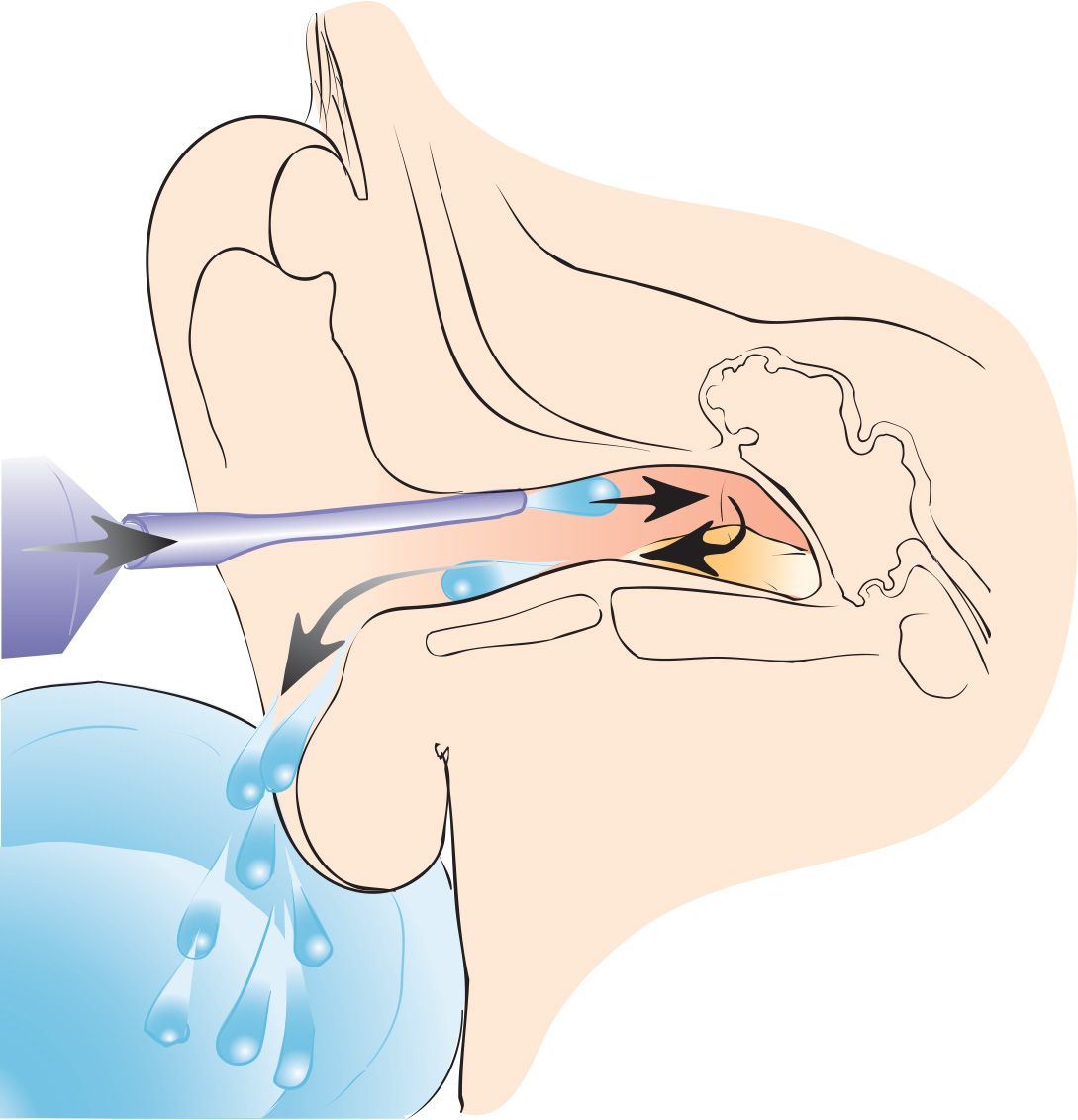
FIGURE 86.2 Syringing cerumen or a foreign body from the auditory canal with an intravenous catheter attached to a 20-mL syringe. The irrigating stream is directed at the posterior, superior wall of the canal. (From Fuerst RS. Removal of cerumen impaction. In: Henretig FM, King C, eds. Textbook of Pediatric Emergency Procedures. Philadelphia, PA: Williams & Wilkins; 1997:643, with permission.)
Related posts:



Stay updated, free articles. Join our Telegram channel

Full access? Get Clinical Tree


