![]() Used to provide local anesthesia to the digits for repair, reduction, or drainage
Used to provide local anesthesia to the digits for repair, reduction, or drainage
![]() Lacerations
Lacerations
![]() Nail bed injuries
Nail bed injuries
![]() Infections (i.e., felons, paronychias)
Infections (i.e., felons, paronychias)
![]() Amputations
Amputations
![]() Fractures or dislocations
Fractures or dislocations
CONTRAINDICATIONS
![]() Absolute Contraindications
Absolute Contraindications
![]() Transthecal technique contraindicated in cases of infection, including felon, tenosynovitis, and overlying cellulitis
Transthecal technique contraindicated in cases of infection, including felon, tenosynovitis, and overlying cellulitis
![]() Allergy to lidocaine, bupivacaine, or other selected anesthetic
Allergy to lidocaine, bupivacaine, or other selected anesthetic
![]() Relative Contraindications
Relative Contraindications
![]() Complex laceration or other injury involving multiple digits that can be more easily and adequately anesthetized with a nerve block at the wrist
Complex laceration or other injury involving multiple digits that can be more easily and adequately anesthetized with a nerve block at the wrist
RISKS/CONSENT ISSUES
![]() Pain (site of needle insertion)
Pain (site of needle insertion)
![]() Bleeding (local at needle puncture site)
Bleeding (local at needle puncture site)
![]() Infection (theoretical risk of iatrogenic infection)
Infection (theoretical risk of iatrogenic infection)
![]() Potential for damage to neurovascular bundle
Potential for damage to neurovascular bundle
![]() Paresthesias
Paresthesias
![]() Possible need for additional anesthetic or alternate procedures if the initial nerve block fails
Possible need for additional anesthetic or alternate procedures if the initial nerve block fails
![]() General Basic Steps
General Basic Steps
![]() Aseptic technique
Aseptic technique
![]() Choose approach and deliver anesthetic
Choose approach and deliver anesthetic
![]() Massage area for 25 to 30 seconds
Massage area for 25 to 30 seconds
![]() Test for adequate analgesia
Test for adequate analgesia
LANDMARKS
![]() The common digital nerves divide into two pairs of nerves corresponding to the dorsal and volar sides of the digits
The common digital nerves divide into two pairs of nerves corresponding to the dorsal and volar sides of the digits
![]() Palmar Nerve
Palmar Nerve
![]() Located at the 4- and 8 o’clock positions when looking at a cross section of the digit
Located at the 4- and 8 o’clock positions when looking at a cross section of the digit
![]() Supplies the volar surface of the digit and the dorsal surface distal to the distal interphalangeal (DIP) joint for the middle three fingers
Supplies the volar surface of the digit and the dorsal surface distal to the distal interphalangeal (DIP) joint for the middle three fingers
![]() Blocking only the palmar nerves will provide adequate anesthesia on fingertip injuries distal to the DIP for the three middle fingers
Blocking only the palmar nerves will provide adequate anesthesia on fingertip injuries distal to the DIP for the three middle fingers
![]() Digital Nerve
Digital Nerve
![]() Located at the dorsal 2- and 10 o’clock positions when looking at a cross section of the digit
Located at the dorsal 2- and 10 o’clock positions when looking at a cross section of the digit
![]() Supplies the nail beds of the thumb, fifth digit, and dorsal aspects of all three middle fingers up to the DIP
Supplies the nail beds of the thumb, fifth digit, and dorsal aspects of all three middle fingers up to the DIP
![]() For the thumb and fifth digit, all four nerves must be blocked for fingertip and nail bed anesthesia (FIGURE 52.1)
For the thumb and fifth digit, all four nerves must be blocked for fingertip and nail bed anesthesia (FIGURE 52.1)
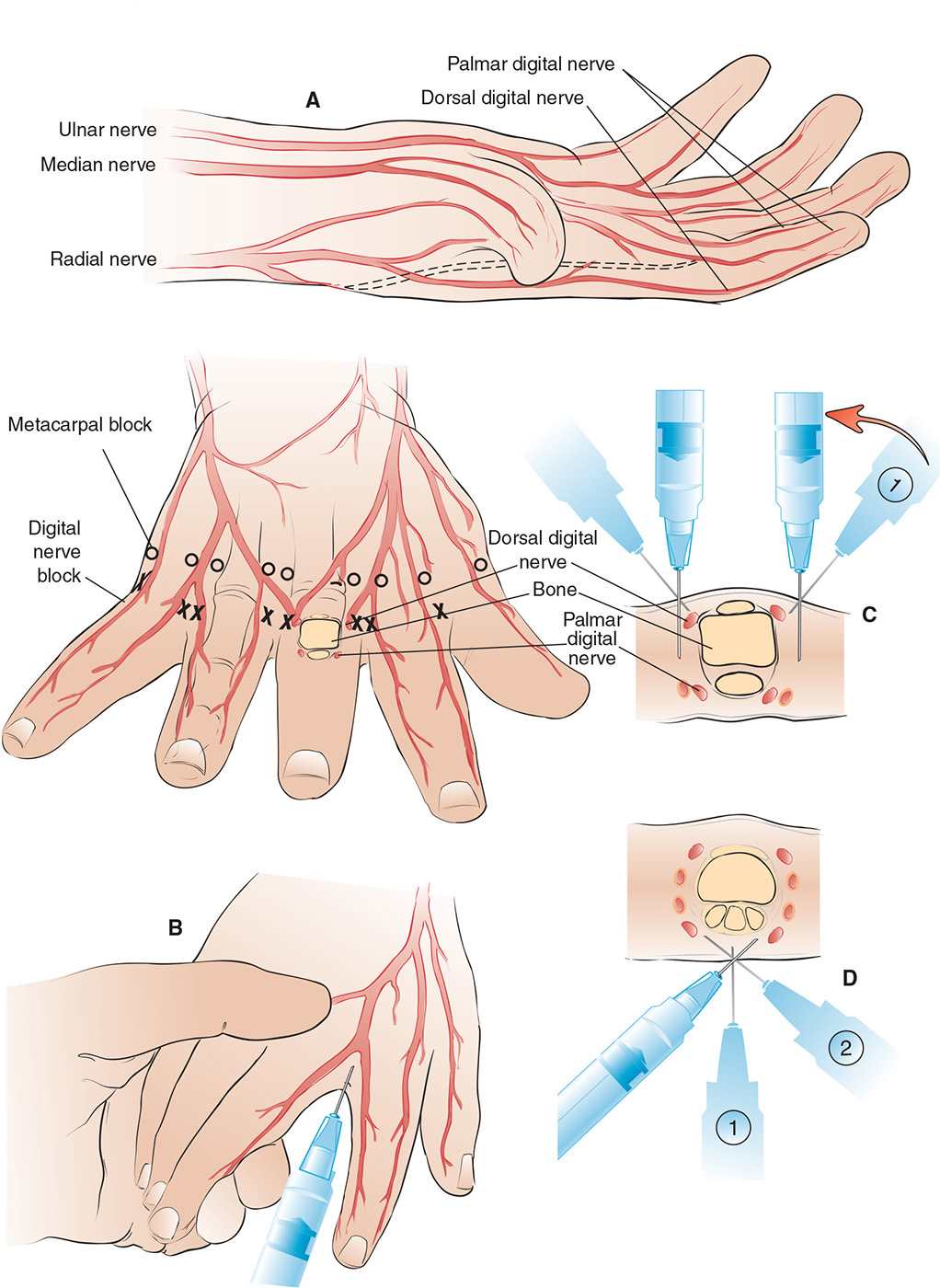
FIGURE 52.1 Dorsal technique for palmar and dorsal digital nerve block. A: Nerve distribution in hand. B: Traditional digital nerve block. C: Dorsal three-sided ring block. D: Volar three-sided ring block. (Lewis L, Stephan M. Local and regional anesthesia. In: Henretig FM, King C, eds. Textbook of Pediatric Emergency Procedures. Philadelphia, PA: Williams & Wilkins; 1997:481, with permission.)
TECHNIQUE: SEVEN APPROACHES
![]() Patient Preparation
Patient Preparation
![]() Document neurovascular examination before anesthesia
Document neurovascular examination before anesthesia
![]() Place patient’s affected hand/foot comfortably on bedside procedure table with volar surface down (for metacarpal nerve block, traditional ring block, wing block) or volar surface up (for subcutaneous block, transthecal approach or thumb block)
Place patient’s affected hand/foot comfortably on bedside procedure table with volar surface down (for metacarpal nerve block, traditional ring block, wing block) or volar surface up (for subcutaneous block, transthecal approach or thumb block)
![]() Prepare the digit and web space by using standard aseptic technique
Prepare the digit and web space by using standard aseptic technique
![]() Equipment
Equipment
![]() Lidocaine or procaine 1% to 2% (or 0.25% bupivacaine for longer, complicated procedures), 2 to 3 mL
Lidocaine or procaine 1% to 2% (or 0.25% bupivacaine for longer, complicated procedures), 2 to 3 mL
![]() An 18-gauge needle for drawing up the anesthetic
An 18-gauge needle for drawing up the anesthetic
![]() A 25- to 30-gauge needle for the nerve block
A 25- to 30-gauge needle for the nerve block
![]() A 5-mL syringe
A 5-mL syringe
![]() Povidone–iodine or chlorhexidine solution
Povidone–iodine or chlorhexidine solution
![]() Sterile drapes and sterile gauze
Sterile drapes and sterile gauze
![]() Gloves
Gloves
![]() Traditional Digital Block (Web-space Block or Metacarpal Nerve Block)
Traditional Digital Block (Web-space Block or Metacarpal Nerve Block)
![]() Anesthetizes all digits except great toe
Anesthetizes all digits except great toe
![]() Prepare skin over dorsal surface of web space between metacarpal/metatarsal heads
Prepare skin over dorsal surface of web space between metacarpal/metatarsal heads
![]() Aspirate and inject subcutaneous wheal between metacarpal/metatarsal bones on dorsum of hand/foot 1 to 2 cm proximal to web space
Aspirate and inject subcutaneous wheal between metacarpal/metatarsal bones on dorsum of hand/foot 1 to 2 cm proximal to web space
![]() Slowly advance needle through the wheal toward lateral volar surface of metacarpal/metatarsal head until slight tenting of the volar surface is appreciated
Slowly advance needle through the wheal toward lateral volar surface of metacarpal/metatarsal head until slight tenting of the volar surface is appreciated
![]() Aspirate and then inject 2 mL of anesthetic
Aspirate and then inject 2 mL of anesthetic
![]() Repeat the process on the opposite side of the finger/toe
Repeat the process on the opposite side of the finger/toe
![]() Traditional Three-sided Ring Block
Traditional Three-sided Ring Block
![]() Anesthetizes all digits including the dorsal, medial, and lateral nerve branches of great toe
Anesthetizes all digits including the dorsal, medial, and lateral nerve branches of great toe
![]() Give two injections of lidocaine, one on each side of the digit
Give two injections of lidocaine, one on each side of the digit
![]() Locate dorsal–lateral aspect of proximal phalanx at the web space, just distal to metacarpal/phalangeal (MCP) or metatarsal/phalangeal (MTP) joint
Locate dorsal–lateral aspect of proximal phalanx at the web space, just distal to metacarpal/phalangeal (MCP) or metatarsal/phalangeal (MTP) joint
![]() Advance needle perpendicular to digit until bone is struck, aspirate and slowly inject 0.5 mL of lidocaine to anesthetize the dorsal nerve
Advance needle perpendicular to digit until bone is struck, aspirate and slowly inject 0.5 mL of lidocaine to anesthetize the dorsal nerve
![]() Withdraw needle slightly, then redirect and advance toward volar surface and slowly inject 1 mL of lidocaine
Withdraw needle slightly, then redirect and advance toward volar surface and slowly inject 1 mL of lidocaine
![]() Withdraw needle partially and redirect it medially over dorsal aspect of digit, aspirate and slowly inject lidocaine while withdrawing needle to anesthetize medial and dorsal aspect of digit
Withdraw needle partially and redirect it medially over dorsal aspect of digit, aspirate and slowly inject lidocaine while withdrawing needle to anesthetize medial and dorsal aspect of digit
![]() Withdraw the needle
Withdraw the needle
![]() Repeat procedure on medial side of digit at site of anesthetized skin
Repeat procedure on medial side of digit at site of anesthetized skin
![]() Massage area of infiltrated skin for 15 to 30 seconds to ensure diffusion of the anesthetic
Massage area of infiltrated skin for 15 to 30 seconds to ensure diffusion of the anesthetic
![]() Wait for 5 to 10 minutes to test for efficacy
Wait for 5 to 10 minutes to test for efficacy
![]() Four-sided Ring Block
Four-sided Ring Block
![]() Advantages: Anesthetizes volar side of digits
Advantages: Anesthetizes volar side of digits
![]() Disadvantages: May result in ischemic complications
Disadvantages: May result in ischemic complications
![]() Perform traditional three-sided ring block
Perform traditional three-sided ring block
![]() Locate anesthetized volar–lateral aspect of proximal phalanx at the web space, just distal to MCP or MTP joint
Locate anesthetized volar–lateral aspect of proximal phalanx at the web space, just distal to MCP or MTP joint
![]() Advance needle medially, aspirate and slowly inject 0.5 mL of lidocaine to anesthetize the volar side while withdrawing needle
Advance needle medially, aspirate and slowly inject 0.5 mL of lidocaine to anesthetize the volar side while withdrawing needle
![]() Subcutaneous Block
Subcutaneous Block
![]() Prepare skin over volar surface at proximal skin crease
Prepare skin over volar surface at proximal skin crease
![]() Pinch skin distal to proximal skin crease
Pinch skin distal to proximal skin crease
![]() Insert needle at midpoint of crease, aspirate and inject subcutaneous 1 to 2 mL wheal
Insert needle at midpoint of crease, aspirate and inject subcutaneous 1 to 2 mL wheal
![]() Massage injected area for 15 to 30 seconds to improve diffusion process
Massage injected area for 15 to 30 seconds to improve diffusion process
![]() Transthecal Approach
Transthecal Approach
![]() Advantages: Single injection and low risk of neurovascular bundle injury
Advantages: Single injection and low risk of neurovascular bundle injury
![]() Disadvantage: More painful to inject through volar surface
Disadvantage: More painful to inject through volar surface
![]() Anesthetic is infused directly into the flexor tendon sheath at the proximal digital crease on volar surface
Anesthetic is infused directly into the flexor tendon sheath at the proximal digital crease on volar surface
![]() Fill 5-mL syringe with lidocaine
Fill 5-mL syringe with lidocaine
![]() Insert 25-gauge needle at a 90-degree angle at the midpoint of the proximal digital crease and advance until bone is struck
Insert 25-gauge needle at a 90-degree angle at the midpoint of the proximal digital crease and advance until bone is struck
![]() Withdraw needle approximately 2 to 3 mm (should be in flexor tendon sheath) and redirect at a 45-degree angle to the long axis of the digit
Withdraw needle approximately 2 to 3 mm (should be in flexor tendon sheath) and redirect at a 45-degree angle to the long axis of the digit
![]() Aspirate and inject 1.5 to 3 mL lidocaine while palpating tendon sheath with other hand; continue until resistance is felt
Aspirate and inject 1.5 to 3 mL lidocaine while palpating tendon sheath with other hand; continue until resistance is felt
![]() After removing the needle, apply pressure over the tendon proximally to facilitate distal spread
After removing the needle, apply pressure over the tendon proximally to facilitate distal spread
![]() Wait for 2 to 3 minutes to test for efficacy of anesthesia
Wait for 2 to 3 minutes to test for efficacy of anesthesia
![]() Most effective for middle three fingers (FIGURE 52.2)
Most effective for middle three fingers (FIGURE 52.2)
![]() Thumb Block
Thumb Block
![]() All four digital nerves must be blocked for complete anesthesia of the thumb
All four digital nerves must be blocked for complete anesthesia of the thumb
![]() Locate the flexor pollicis longus on the volar aspect of the thumb at the level of the proximal thumb flexor crease
Locate the flexor pollicis longus on the volar aspect of the thumb at the level of the proximal thumb flexor crease
![]() The nerves lie immediately adjacent to this tendon
The nerves lie immediately adjacent to this tendon
![]() Aspirate and inject 1 to 2 mL of lidocaine along both sides of the tendon (FIGURE 52.3)
Aspirate and inject 1 to 2 mL of lidocaine along both sides of the tendon (FIGURE 52.3)
![]() Wing Block
Wing Block
![]() Anesthetizes distal digit and nail bed
Anesthetizes distal digit and nail bed
![]() Prepare the distal digit by using standard aseptic technique
Prepare the distal digit by using standard aseptic technique
![]() Insert 30-gauge needle perpendicular to the long axis of digit at a 45-degree angle at a point 3 mm proximal to the imaginary intersection of the lateral and proximal nail folds
Insert 30-gauge needle perpendicular to the long axis of digit at a 45-degree angle at a point 3 mm proximal to the imaginary intersection of the lateral and proximal nail folds
![]() Aspirate and inject anesthetic across the dorsum of digit parallel to proximal nail fold
Aspirate and inject anesthetic across the dorsum of digit parallel to proximal nail fold
![]() Partially withdraw needle and redirect along lateral nail fold
Partially withdraw needle and redirect along lateral nail fold
![]() Aspirate and inject anesthetic along the lateral nail fold
Aspirate and inject anesthetic along the lateral nail fold
![]() Repeat procedure on opposite side of digit if bilateral anesthesia is required (FIGURE 52.4)
Repeat procedure on opposite side of digit if bilateral anesthesia is required (FIGURE 52.4)
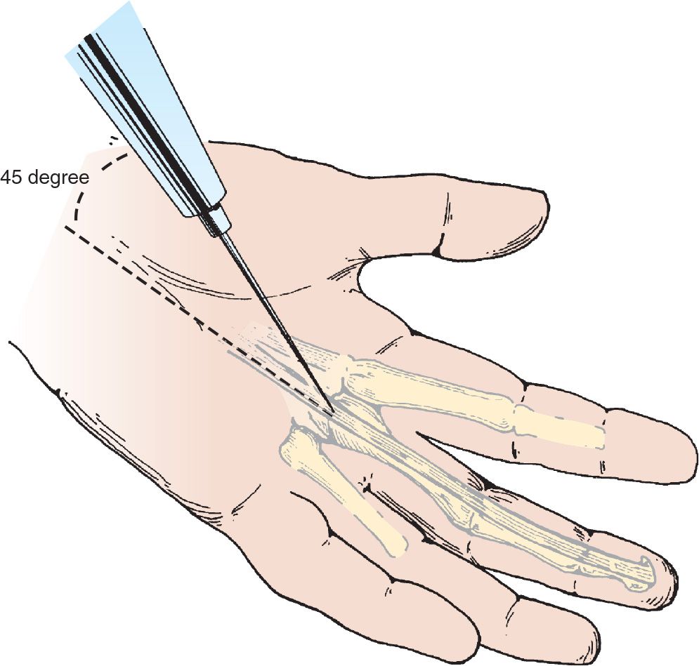
FIGURE 52.2 Digital nerve block: Transthecal approach. The needle is directed into the proximal digital crease at a 45-degree angle to the long axis of the digit into the flexor tendon sheath where the lidocaine is deposited slowly.
Related posts:
Stay updated, free articles. Join our Telegram channel

Full access? Get Clinical Tree


