Continuous Sciatic Blocks
A. Parasacral Approach
Elizabeth Gaertner
blood. The catheter is introduced 3 to 5 cm beyond the needle tip. The introducer needle is removed, and the catheter is secured in place with Steri-Strip (3M, St. Paul, MN) and covered with a transparent dressing.
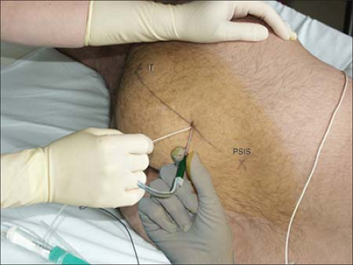 Figure 29-1. The insulated Tuohy needle connected to a nerve stimulator is introduced perpendicularly. |
The site of the introduction of the needle is just below the PSIS.
A motor response at the level of the hip or thigh should not be considered sufficient because it can be produced by direct muscle stimulation or can correspond to a nerve stimulation of the piriformis or the obturator internus muscle (needle too lateral). Conversely, a stimulation of the obturator nerve (adduction of the thigh) indicates that the needle is too anterior and medial. Finally, a gluteal muscle contraction indicates that the position of the needle is too superficial.
In case of bony contact (sacral or iliac bone, near the sacroiliac joint, at the top of the greater sciatic notch), the needle needs to be withdrawn and reintroduced more caudally on the PSIS–IT line. The depth at which the bone was contacted is an important indicator. Since the nerve is approached on the top of the greater sciatic foramen, where the sciatic nerve leaves the pelvis, the needle tip should be introduced no more than an additional 20 mm.
This block is often associated with a block of the obturator nerve (>90%) because of the anatomic proximity of the sacral plexus and the obturator nerve. An extension of the block to the pudendal plexus is also possible, especially to the pudendal nerve (up to 80%). This explains the associated unilateral anesthesia of the perineum. Finally, the proximity of the pelvic splanchnic nerves and the terminal branches of the sympathetic trunks and the inferior hypogastric plexus may explain associated urinary retention.
Although no specific serious complications have been reported with this technique, caution should be exercised to avoid damaging the pelvic vessels and organs because of the proximity of the sacral plexus and the pelvic vessels and organs.
In the recovery room, a radiograph may be performed after injection of a contrast agent to verify the position of the parasacral catheter.
This is the only approach to the sciatic nerve that allows consistent blocking of the tibial, common peroneal, and posterior cutaneous nerves of the thigh and also the superior and inferior gluteal nerves and the quadratus femoris nerve.
Compared with a single parasacral block, the Tuohy needle is oriented caudally and laterally to facilitate the placement of the catheter.
Suggested Readings
Birnbaum K, Preschera A, Hessler S, et al. The sensory innervation of the hip joint—an anatomical study. Surg Radiol Anat 1997;19:371–375.
Bruell P. Sciatic nerve block: parasacral approach. Reg Anesth Pain Med 1998;23:78.
Cuvillon P, Ripart J, Jeannes P, et al. Comparison of the parasacral approach and the posterior approach, with single- and double-injection techniques, to block the sciatic nerve. Anesthesiology 2003;98(6):1436–1441.
Jochum D, Iohom G, Choquet O, et al. Adding a selective obturator nerve block to the parasacral sciatic block: an evaluation. Anesth Analg 2004;99:1544–1549.
Mansour NY, Bennetts FE. An observational study of combined continuous lumbar plexus and single shot sciatic nerve blocks for post-knee surgery analgesia. Reg Anesth 1996;21:287–291.
Morris GF, Lang SA, Dust WN, et al. The parasacral sciatic nerve block. Reg Anesth 1997;22: 223–228.
Ripart J, Cuvillon P, Nouvellon E, et al. Parasacral approach to block the sciatic nerve: a 400-case survey. Reg Anesth Pain Med 2005;30:193–197.
B. Gluteal Approaches
Rama Joshi
1. Gluteal Approach
Related posts:


Stay updated, free articles. Join our Telegram channel

Full access? Get Clinical Tree


