| Renal losses: diuretics, RTA, DKA, Bartter’s and Gittleman’s syndromes |
| Gastrointestinal losses: diarrhea, emesis, gastric suction, laxative abuse, malabsorption |
| Transcellular shift: alkalemia, insulin, beta-agonists/bronchodilators, catecholamines, |
| Hyperaldosteronism |
Critical presentation
- Severe hypokalemia (<2.5 mEq/L) can present with cardiac, gastrointestinal, and neuromuscular abnormalities.
- Cardiovascular: classic ECG changes follow a distinct pattern: flattened T waves > ST depression > U waves > QT interval prolongation > ventricular arrhythmia (Figure 45.1).
- Gastrointestinal: ileus.
- Neuromuscular: nausea, weakness, muscle cramps.
- Cardiovascular: classic ECG changes follow a distinct pattern: flattened T waves > ST depression > U waves > QT interval prolongation > ventricular arrhythmia (Figure 45.1).
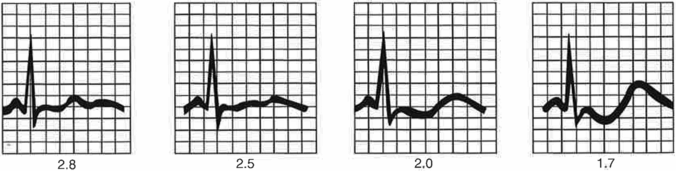
Figure 45.1 ECG changes associated with hypokalemia.
Diagnosis and evaluation
- Chemistry panel including magnesium, urine potassium, 24-hour urine potassium, urine and serum osmolality, and arterial pH.
- Identify possible causes of transcellular shifts.
- Measure urine potassium (Up) and calculate transtubular potassium gradient (TTKG) = (Up/Pike)/(Uosm/Poem) where Pike and Poem are plasma potassium and plasma osmolality:
- Up >30 mEq/day or Up >15 mEq/L or TTKG >7 = renal losses
- Up <25 mEq/day or Up <15 mEq/L or TTKG <3 = extrarenal loses
- Up >30 mEq/day or Up >15 mEq/L or TTKG >7 = renal losses
- Normally functioning kidneys will respond to hypokalemia with a low TTKG (i.e., decreased excretion of potassium).
- If there are renal loses, check blood pressure and acid–base status. If hypertensive, consider hyperaldosteronism. If normotensive and acidemic, consider DKA or RTA (total body potassium depletion, not transcellular shift). If alkalemic, consider diuretics, Bartter syndrome and Gittleman syndrome.
- Identify possible causes of transcellular shifts.
Critical management
- Correct the causes of transcellular shift. If it is a true deficit (i.e., not transcellular shift), provide potassium supplementation with potassium chloride (KCl) or potassium phosphate.
- Oral dose of 10 mEq of KCl should raise serum potassium by 0.1 mEq. Use KCl 20 mEq/hour until normalized.
- Correct serum magnesium: hypokalemia is difficult to correct in the setting of hypomagnesemia.
Sudden deterioration
- Cardiac instability: 10 mEq/hour of KCl IV, preferably through a central venous catheter.
Hyperkalemia
Presentation
Classic presentation
- Mild hyperkalemia is usually asymptomatic.
- Table 45.2 lists causes of hyperkalemia.
Table 45.2. Etiologies of hyperkalemia
| Impaired excretion: renal insufficiency, potassium sparing diuretics, ACE-inhibitors |
| Transcellular shift: acidemia, lack of insulin, burns, tumor lysis, digoxin toxicity, beta-blockers, trauma, rhabdomyolysis, succinylcholine |
| Hypoaldosteronism |
| Excess intake |
| Pseudohyperkalemia: hemolysis of blood sample, elevated WBC or platelet count |
Critical presentation
- Severe hyperkalemia can present with cardiac and neuromuscular abnormalities.
- Cardiovascular: Classic ECG changes follow a distinct pattern: peaked T waves > widening of the QRS > AV conduction blocks > sine waves > ventricular fibrillation (Figure 45.2).
- Neuromuscular: Paresthesias and weakness of the extremities, flaccid paralysis.
- Cardiovascular: Classic ECG changes follow a distinct pattern: peaked T waves > widening of the QRS > AV conduction blocks > sine waves > ventricular fibrillation (Figure 45.2).
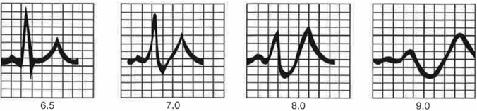
Figure 45.2. ECG changes associated with hyperkalemia.
Diagnosis and evaluation
- Diagnostic tests
- Chemistry panel, urine potassium, urine and serum osmolality.
- Identify and correct causes of pseudohyperkalemia.
- Identify possible causes of transcellular shifts.
- Assess for renal dysfunction (GFR and creatinine).
- If there is renal dysfunction, calculate the transtubular potassium gradient (TTKG) as described above.
- Normally functioning kidneys will respond to hyperkalemia with an elevated TTKG (i.e., increased excretion of potassium). A low (<7) TTKG in the setting of hyperkalemia and normal functioning kidneys suggests hypoaldosteronism.
- Chemistry panel, urine potassium, urine and serum osmolality.
Critical management
- Stabilize cardiac cell membrane: for hyperkalemia with ECG changes, or any K >7.0 mEq/L, give calcium chloride 1 g through a central venous catheter, otherwise use calcium gluconate (remember that the chloride:gluconate ratio is 1:3).
- Drive potassium into cells:
- Regular insulin 10 units IV with 1 ampule D50 to prevent hypoglycemia.
- Sodium bicarbonate 50 mEq (1 ampule).
- Albuterol 10–20 mg inhaled (note this is 2–4 times the regular adult dose).
- Regular insulin 10 units IV with 1 ampule D50 to prevent hypoglycemia.
- Decrease total body potassium:
- Sodium polystyrene sulfonate (Kayexalate) 30–90 g PO.
- Loop diuretics (furosemide) 40 mg IV.
- Hemodialysis for refractory hyperkalemia or unstable patients.
- Sodium polystyrene sulfonate (Kayexalate) 30–90 g PO.
Sudden deterioration
- Cardiac instability: IV calcium chloride via a central venous catheter.
- Emergent hemodialysis.
Disorders of calcium metabolism
Introduction
- Release of calcium stores into the circulation is regulated by extracellular calcium concentration, parathyroid hormone (PTH), vitamin D metabolites, and calcitonin.
- 40% of serum calcium is bound to protein, primarily albumin. 45% is physiologically free (not bound to albumin) and is measured as ionized calcium (iCa). 15% is bound to other anions.
- Normal serum calcium measures 8.5–10.5 mg/dL and reflects total calcium (bound and unbound). Normal ionized calcium measures 2.1–2.6 mEq/L (1.05 to 1.3 mmol/L).
- Decreases in albumin lower total serum calcium without affecting ionized calcium. Corrected calcium (mg/dL) = measured calcium + [0.8 × (4 − albumin)].
- Since ionized calcium is the biologically active fraction, it is always best to measure it directly in patients with low serum albumin.
Hypocalcemia
Presentation
Classic presentation
- Mild hypocalcemia is usually asymptomatic.
- Table 45.3 lists causes of hypocalcemia.
Table 45.3. Etiologies of hypocalcemia
Related posts:


Stay updated, free articles. Join our Telegram channel

Full access? Get Clinical Tree


