Fig. 17.1
A lethal danger spot in right colon dissection: Henle’s gastrocolic trunk (Ignjatovic et al. 2004; Lange et al. 2000). (a) Demonstration of the gastrocolic trunk of Henle (GTH) with the corrosion cast method. ASPDV, anterior superior pancreaticoduodenal vein; GTH, gastrocolic trunk of Henle; JV, jejunal vein (prima); MCV, middle colic vein; RGEV, right gastroepiploic vein; SMA, superior mesenteric artery; SMV, superior mesenteric vein; SV, splenic vein. (b) Variations of Henle’s gastrocolic trunk: the anatomy of venous tributaries of the superior mesenteric vein at the inferior border of the pancreas. Numbers indicate numbers of subjects A, Superior mesenteric vein; B, right gastroepiploic vein; C, anterior superior pancreaticoduodenal vein; D, right superior colic vein
The only reasonable suggestion is to be gentle and avoid this injury.
Hemostatic clamps and stitches complete dissection at this end of the gastrocolic ligament.
17.3.2 Transverse Colon
Omental resection is optional.
Particular attention should be paid not to tear the splenic capsule when dissecting near the splenic flexure and/or while ligating the left end of the gastrocolic ligament and/or splenocolic attachments.
Mobilization (without resection) of the spleen may facilitate this dissection, as well as a surgical swab placed gently above spleen and below diaphragm.
A distended megacolon, or an inflammatory, diseased colon, is vulnerable to tears and/or perforation at or near the splenic flexure.
17.3.3 Left Descending Colon
The nondominant hand elevates the colon, extracting it out of the abdomen and to the patient’s right, while the assistant retracts the abdominal wall to the left. In this manner, the white (Toldt) line comes into view under tension between the parietal and descending colon peritoneums. Cutting with cautery precisely on this line exposes the underlying alveolar tissue. Gentle traction and cautery free the descending colon, which now is attached only by its mesentery.
17.3.4 Sigmoid
The sigmoid root has a length of 5–10 cm; the sigmoid mesentery unfolds like a fan to 25–60 cm. The surgeon’s and assistant’s positions as well as traction are similar to those followed in descending colonic mobilization; the sigmoid is mobilized by continuing the cautery incision on Toldt’s line at the outer aspect of the sigmoid. Alveolar tissue is exposed and can be pushed with a wet sponge down to the root of the sigmoid mesentery, care being taken to avoid injury to the left spermatic vessels, and visualizing the left ureter. The ureter lies on the posterior abdominal wall, crossing anteriorly the bifurcation of the internal and external iliac vessels. The ureter contracts with a gentle touch of an atraumatic instrument; no need to mark or tape it, just identify it to make sure to avoid it.
17.3.5 Rectum
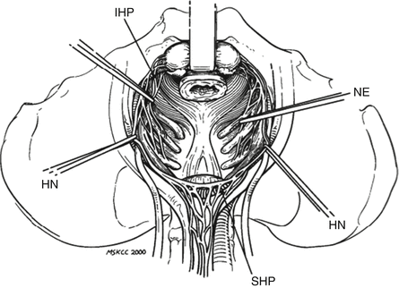
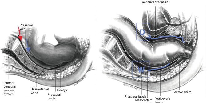
As the sigmoid is pulled out of the abdomen, the peritoneal surface at the medial aspect of the mesosigmoid root is incised, from the aortic bifurcation caudally along the medial aspect of the right iliac vessels, and the incision is continued between the rectum and pelvic brim, rectum and bladder or uterus, as the assistant applies opposite traction to these organs. Parallel and superficial to the aortic bifurcation lie the hypogastric nerves (sexual function) (Fig. 17.2). Once identified, avoid traction on the nerves during the next step. Following the alveolar plane below the aortic bifurcation bluntly down to the pelvic cavity, the rectum/mesorectum can be dissected free from the presacral space, down to Waldayer’s fascia (Fig. 17.3). If cancer is not the problem, mobilization is accomplished within seconds by gentle insertion of the dominant hand. Neither cautery nor ligation is needed. Avoid pressing against mesorectum with the tip of fingers, because this may perforate a fragile rectum, leading to troublesome bleeding and a source of potential contamination; use the palm of the hand. To complete rectal mobilization circumferentially, using (long shaft) cautery bursts dissect all connective tissues laterally from both sides freeing the lateral mesorectum from the pelvic fascia. Usually, no vessel is encountered: no need for ligation, as simple cautery forceps suffice. Finally the anterior rectal plane is incised and freed from its attachments to the uterus/vagina in women or bladder/seminal vesicles/prostate in men. Putting tension to rectum by posterior traction and contra-tension by pulling (e.g., with a St. Marks-type retractor), the anterior tissues (vagina, bladder, etc.) against the pubic bone, the correct plane is found (Fig. 17.3); it is essential to remain in the specific plane (Denonvillier’s fascia) until reaching the deepest part of dissection, avoiding damage on nervi erigentes and its branches, responsible for sexual function (Fig. 17.2). In women, vision can sometimes be improved and working space increased by temporarily stitching the dome of the uterus to the pubic skin, elevating it out of the operating field.


Fig. 17.2
The hypogastric plexus. Dissecting in the correct pelvic plane (see Fig. 17.3) preserves nerve and sexual function. IHP inferior hypogastric nerve plexus, NE nervi erigentes, HN hypogastric nerve, SHP superior hypogastric nerve plexus

Fig. 17.3
Lateral pelvis view. The correct plane to start on mesorectum (left image, blue arrow) and continue the dissection (right image, blue arrows); avoids hemorrhage, nerve damage, injury to adjacent organs. Waldayer’s and Denonvilier’s fascias. False plane of pelvic dissection (Red arrow)
17.4 Vessel Ligation
Vessel ligation differs according to the disease and what segment (right and left colectomy, sigmoidectomy, low anterior resection, or segmental resection) is performed. The regional lymph nodes reside along the feeding vessels and can be removed as needed. In a non-oncologic emergency, just the diseased part of the colon along with a sphenoid part of its mesentery is all that has to be excised. The appex of this sphenoid part goes down to the mesenteric root, so there are fewer vessels to ligate, saving time. Energy-driven devices (which seal and cut vessels) are effective especially in areas with diminished working space (i.e., pelvis) and save time.
17.4.1 Right Hemicolectomy
Right hemicolectomy: Having mobilized the right colon the vessels to ligate include the ileocolic, right colic, and right branch of middle colic (retain main stem and the left branch) arteries.
17.4.2 Left Hemicolectomy
Ligation of the inferior mesenteric at its origin (extended left hemicolectomy) means that adequate vascular supply relies on the marginal artery (from the transverse colon) above, and requires excision of the sigmoid and extraperitoneal rectum with the specimen.
17.4.3 Sigmoidectomy
Sigmoid trunk ligation at the root usually suffices. The taenia coli disappear at the rectosigmoid juncture, forming a complete outer muscular layer in the upper rectum, which in addition is the narrowest part of the colonic lumen. This anatomic characteristic has been incriminated as responsible for diverticular disease, and is the rationale for mandatory resection of this portion in diverticular disease. Ligation of the superior rectal artery (continuation of the inferior mesenteric artery) is not mandatory, and some advise its preservation.
17.4.4 Low Anterior Rectal Resection
In low anterior rectal resection (defined as resection of the proximal two thirds of the rectum, leaving the sphincter mechanism intact, and anastomosis below peritoneal reflection) superior, middle, and inferior rectal arteries are ligated depending on the depth of resection of the rectum. Middle and inferior arteries are rarely visualized and safely sealed with cautery, or energy-driven devices. Retaining the rectal ampulla or an ileorectal anastomosis (for total colectomy) are important for quality of life.
17.5 Anastomoses
Prerequisites include:
1.
Good blood supply to the bowel margins: this can be evaluated by the color of the divided bowel wall (in comparison with the adjacent distal colon), ample bleeding at the cut edge (or an nearby epiploic appendix), and also by Doppler and/or indocyanine green.
2.
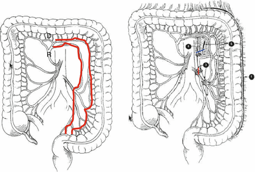
Avoid tension: a rule of thumb is that the two extremities must overlap each other for at least 2 cm, without traction in an end to end anastomosis. If not, further mobilization and/or mesenteric incisions are mandatory, even if it means occasionally sacrificing a major vessel. In a low rectal anastomosis where the rectum cannot be mobilized, further length should come from the descending colon (splenic flexure mobilization) with ligation and division of the inferior mesenteric vein under the pancreas along the ligament of Treitz, and the inferior mesenteric artery at its root, leaving intact the bypassing branches of the marginal artery of Drummond and the arc of Riolan supplying blood from the superior mesenteric artery (Fig. 17.4). Always be cautious when there has been previous operations (that have potentially occluded the arterial arcades), radiation therapy, atherosclerosis and diabetes, or even radical nephrectomy. Check colonic viability before ligating the main vessels by temporary vessel occlusion with a bulldog.


Fig. 17.4
Anastomotic arterial arc of Riolan, and marginal artery or Drummond, are the feeding arteries of a long descending colon graft formed after division of inferior mesenteric artery (red line) and vein (blue line) (right image)
3.
Once completed, the anastomosis should be visually and palpably evaluated for tension. It should lie gently on the surroundings.
4.
If a tension-less anastomosis is not possible, create a stoma or, if an anastomosis is already created, add a prophylactic ileostomy. Drains or delaying the patient’s oral feeding will not heal an unsafe anastomosis.
Technical issues regarding anastomoses:
Hand sewn: Hand sewn or stapled anastomoses can be performed according to personal preferences: there is no significant differences in leakage rates; however, the immediate risks of bleeding (should decrease with new multi (>2) staple lines) and the long-term risk of stricture are higher with the staples. Speed of construction depends on the operator, more than on the method. One layer, ideally extramucosal, is enough. Interrupted or continuous is also a matter of surgeons’s preference and provide equally satisfactory results when well done.
Stay updated, free articles. Join our Telegram channel

Full access? Get Clinical Tree


