Fig. 24.1
Type of infective disease according to anatomical layer
):
1.
Skin
2.
Subcutaneous tissue
3.
Muscles (separated from subcutaneous tissue by fascia or aponeurosis)
Three anatomic layers correspond to three types of infective diseases:
1.
Infectious dermatitis
2.
Subcutaneous gangrene
3.
Myositis
Nota bene: Subcutaneous tissue is frequently referred to as “superficial fascia” and aponeurosis to “deep fascia.” These terms are confusing as is the term “fasciitis”: Aponeurosis is a barrier to spread of infection and should be preserved when treating subcutaneous gangrene.
24.1 Dermatitis
Infectious dermatitis may be caused by:
Gram+ cocci alone
Impetigo
Ecthyma
Erysipelas
Several anaerobic and anaerobic bacteria, Gram+ and Gram – acting in synergy
Fournier’s gangrene
24.1.1 Impetigo
Highly contagious pyoderma
Children younger than 6 years most frequently affected
Primarily caused by Staphylococcus aureus (sometimes by Streptococcus pyogenes)
Painless lesions on the face, scalp, arms, and legs
Begins as vesicles evolving to pustules then to honey-colored crusts
Usually without associated general signs of infection
24.1.2 Ecthyma
Ulcerative pyoderma.
Caused by Staphylococcus aureus or by Streptococcus pyogenes.
May be caused by Pseudomonas.
Different stages of lesions may coexist.
Painful.
Begins as a pustule that evolves into a deep ulcer covered by a crust.
Satellite lymph nodes are swollen.
Heals, leaving definitive scars.
24.1.3 Pyoderma Gangrenosum
Not an infection
Results from an immune system dysfunction as in inflammatory bowel disease, myeloma, or rheumatoid arthritis.
Begins as a papule, followed by a vesicle ending as a necrotic ulcer.
Different stages of lesions may coexist.
Treated by corticoids.
24.1.4 Erysipelas
Acute streptococcal dermatitis.
Associated with general illness and fever.
Erythematous skin lesions enlarge rapidly.
Sharply demarcated raised edge.
Red streak and swollen lymph nodes may be present.
24.1.5 Fournier’s Gangrene
All gangrene of the genitalia are not Fournier’s gangrene.
Caused by mixed aerobic and anaerobic bacteria.
Begins suddenly with fever and deep general illness.
Inoculation occurs after a wound or operation.
Lesion consists of superficial skin necrosis, typically (but not always) with border between necrotic and healthy skin.
Crepitus may be present.
Necrosis heals as a deep second-degree burn.
Surgery is required to remove necrotic tissues.
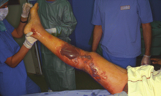
24.2 Subcutaneous Gangrene (SG)
24.2.1 Hemolytic Streptococcus Subcutaneous Gangrene (HSSG)
This disease was described by F.L. Meleney in 1924 as “haemolytic streptococcal gangrene.” The subsequent terms of suppurative or necrotizing fasciitis must be avoided, as they are confusing:
the deep fascia or aponeurosis is a barrier that generally prevents deeper extension of the infection.
Caused by a Streptococcus pyogenes group A, B, C, or G inoculated through a skin wound.
General illness is profound.
Red swollen area with no demarcated edge (contrary to erysipelas) (Fig. 24.2).