![]() A Colles fracture is a transverse fracture through the distal 2 to 3 cm of the radial metaphysis where the distal fragment is dorsally displaced and angulated. The most common mechanism is a fall on an outstretched hand.
A Colles fracture is a transverse fracture through the distal 2 to 3 cm of the radial metaphysis where the distal fragment is dorsally displaced and angulated. The most common mechanism is a fall on an outstretched hand.
![]() Closed reduction is indicated if distal fragment has a dorsal tilt >10 degrees, an intra-articular fracture is present and has a >1 mm step-off, or there is >2 mm radial shortening
Closed reduction is indicated if distal fragment has a dorsal tilt >10 degrees, an intra-articular fracture is present and has a >1 mm step-off, or there is >2 mm radial shortening
![]() General goals are to reduce displaced fragments and maintain reduction during healing
General goals are to reduce displaced fragments and maintain reduction during healing
CONTRAINDICATIONS
![]() Hematoma block contraindicated if:
Hematoma block contraindicated if:
![]() History of allergy to local anesthetics
History of allergy to local anesthetics
![]() Overlying skin infection or dirty skin
Overlying skin infection or dirty skin
![]() Reduction contraindicated if open fracture exists
Reduction contraindicated if open fracture exists
PROCEDURAL RISKS/CONSENT ISSUES
![]() Pain (site of needle insertion)
Pain (site of needle insertion)
![]() Bleeding (local at needle puncture site)
Bleeding (local at needle puncture site)
![]() Infection (theoretical risk of iatrogenic infection)
Infection (theoretical risk of iatrogenic infection)
![]() General Basic Steps
General Basic Steps
![]() Obtain radiographs
Obtain radiographs
![]() Hematoma block
Hematoma block
![]() Reduction
Reduction
![]() Splinting
Splinting
![]() Postreduction steps
Postreduction steps
LANDMARKS: RADIOGRAPHIC
![]() Standard radiographs should include a posteroanterior (PA) and a lateral projection
Standard radiographs should include a posteroanterior (PA) and a lateral projection
![]() Clearly describe fractures as pediatric or adult, extra-articular or intra-articular, comminuted or noncomminuted, angulated or not angulated
Clearly describe fractures as pediatric or adult, extra-articular or intra-articular, comminuted or noncomminuted, angulated or not angulated
![]() In adults, several measurements are used to determine the extent of deformity
In adults, several measurements are used to determine the extent of deformity
![]() Radial height (PA view): Two parallel lines drawn perpendicularly to the long axis of the radius, one through the tip of the radial styloid and the other at the articular surface of the radius
Radial height (PA view): Two parallel lines drawn perpendicularly to the long axis of the radius, one through the tip of the radial styloid and the other at the articular surface of the radius
![]() Normal radial height is 9.9 to 17.3 mm
Normal radial height is 9.9 to 17.3 mm
![]() Radial inclination (PA view): A line drawn through the articular surface of the radius, perpendicular to its long axis. A line is then drawn tangent from the tip of the radial styloid.
Radial inclination (PA view): A line drawn through the articular surface of the radius, perpendicular to its long axis. A line is then drawn tangent from the tip of the radial styloid.
![]() Normal radial inclination is 15 to 25 degrees
Normal radial inclination is 15 to 25 degrees
![]() Volar tilt (lateral view): A line drawn perpendicularly to the long axis of the radius. A line is then drawn tangent to it along the articular surface from the dorsal to palmar surface of the radius.
Volar tilt (lateral view): A line drawn perpendicularly to the long axis of the radius. A line is then drawn tangent to it along the articular surface from the dorsal to palmar surface of the radius.
![]() Normal volar tilt is 10 to 25 degrees
Normal volar tilt is 10 to 25 degrees
SUPPLIES
![]() Povidone–iodine or chlorhexidine solution
Povidone–iodine or chlorhexidine solution
![]() 25-gauge needle and 10- to 20-mL syringe for hematoma block
25-gauge needle and 10- to 20-mL syringe for hematoma block
![]() Local anesthesia: 1% lidocaine without epinephrine or bupivacaine 0.5%
Local anesthesia: 1% lidocaine without epinephrine or bupivacaine 0.5%
![]() Reduction materials: Gauze bandage roll for finger trap, traction weights (8–10 lb)
Reduction materials: Gauze bandage roll for finger trap, traction weights (8–10 lb)
![]() Splinting materials: Web roll, plaster, elastic compression bandage
Splinting materials: Web roll, plaster, elastic compression bandage
TECHNIQUE
![]() Clinical Assessment
Clinical Assessment
![]() Inspection: Identify the skeletal deformity. Classic finding is the so-called dinner-fork deformity, produced by dorsal displacement of the distal fracture fragments.
Inspection: Identify the skeletal deformity. Classic finding is the so-called dinner-fork deformity, produced by dorsal displacement of the distal fracture fragments.
![]() Palpation: Note any step-off, crepitus, and the point of maximal tenderness
Palpation: Note any step-off, crepitus, and the point of maximal tenderness
![]() Test neurovascular status: Acute median nerve compression is common in these injuries, especially in severely displaced, high-energy fractures. Pay close attention to finger sensation.
Test neurovascular status: Acute median nerve compression is common in these injuries, especially in severely displaced, high-energy fractures. Pay close attention to finger sensation.
![]() Evaluate for a distal radioulnar joint (DRUJ) dislocation: Caused by a disruption of the triangular fibrocartilage complex which stabilizes the joint. Orthopedic consultation is necessary for this injury.
Evaluate for a distal radioulnar joint (DRUJ) dislocation: Caused by a disruption of the triangular fibrocartilage complex which stabilizes the joint. Orthopedic consultation is necessary for this injury.
![]() X-rays may be reported as normal; physical examination is the key to diagnosis
X-rays may be reported as normal; physical examination is the key to diagnosis
![]() Wrist has limited range of motion, with crepitus on supination and pronation
Wrist has limited range of motion, with crepitus on supination and pronation
![]() Loss of the ulnar styloid contour with volar ulna dislocation and prominence of the ulnar styloid with dorsal dislocation
Loss of the ulnar styloid contour with volar ulna dislocation and prominence of the ulnar styloid with dorsal dislocation
![]() More frequently with associated ulnar styloid fracture
More frequently with associated ulnar styloid fracture
![]() Evaluate for a Salter–Harris type I fracture in pediatric patients
Evaluate for a Salter–Harris type I fracture in pediatric patients
![]() Tenderness over the distal radial physis
Tenderness over the distal radial physis
![]() Only radiologic finding may be displacement or absence of the pronator quadratus fat pad sign
Only radiologic finding may be displacement or absence of the pronator quadratus fat pad sign
![]() Low threshold to splint and arrange orthopedic follow-up
Low threshold to splint and arrange orthopedic follow-up
![]() Rarely results in a growth disturbance
Rarely results in a growth disturbance
![]() Consider child abuse in patients <1 year of age with this injury (FIGURE 50.1)
Consider child abuse in patients <1 year of age with this injury (FIGURE 50.1)
![]() Hematoma Block
Hematoma Block
![]() Prepare skin over fracture site with povidone–iodine or chlorhexidine solution
Prepare skin over fracture site with povidone–iodine or chlorhexidine solution
![]() Insert a 25-gauge needle dorsally into the hematoma at the fracture site approximately 30 degrees to the skin. Guide the needle tip into the fracture space by sliding along the fractured surface of the distal fragment. Placement is confirmed by the aspiration of blood.
Insert a 25-gauge needle dorsally into the hematoma at the fracture site approximately 30 degrees to the skin. Guide the needle tip into the fracture space by sliding along the fractured surface of the distal fragment. Placement is confirmed by the aspiration of blood.
![]() Slowly inject 5 to 10 mL of 1% lidocaine without epinephrine into the fracture cavity and another 5 mL into the surrounding periosteum
Slowly inject 5 to 10 mL of 1% lidocaine without epinephrine into the fracture cavity and another 5 mL into the surrounding periosteum
![]() Lidocaine will provide anesthesia for approximately 1 to 2 hours
Lidocaine will provide anesthesia for approximately 1 to 2 hours
![]() Bupivacaine 0.5% may also be used if available and has a significantly longer duration of action (4 to 6 hours)
Bupivacaine 0.5% may also be used if available and has a significantly longer duration of action (4 to 6 hours)
![]() Allow 10 to 15 minutes for the anesthesia to become effective
Allow 10 to 15 minutes for the anesthesia to become effective
![]() Reduction (Jones Method): Goal is to restore the normal anatomy (radial height, radial inclination, volar tilt, and intra-articular step-off) through traction and manipulation (FIGURE 50.2)
Reduction (Jones Method): Goal is to restore the normal anatomy (radial height, radial inclination, volar tilt, and intra-articular step-off) through traction and manipulation (FIGURE 50.2)
![]() Place patient’s fingers in a finger trap device with the elbow in 90 degrees of flexion
Place patient’s fingers in a finger trap device with the elbow in 90 degrees of flexion
![]() Suspend 8 to 10 lb of weight from elbow (distal humerus specifically) for 5 to 10 minutes to disimpact fracture fragments
Suspend 8 to 10 lb of weight from elbow (distal humerus specifically) for 5 to 10 minutes to disimpact fracture fragments
![]() While in traction, apply dorsal pressure over the distal fragment with your thumbs while simultaneously applying volar pressure over the proximal segment with your fingers to continue to disimpact the fragments
While in traction, apply dorsal pressure over the distal fragment with your thumbs while simultaneously applying volar pressure over the proximal segment with your fingers to continue to disimpact the fragments
![]() Apply volar force to the distal fragments to realign them into anatomic position
Apply volar force to the distal fragments to realign them into anatomic position
![]() Remove the traction weight
Remove the traction weight
![]() Splinting: A sugar-tong splint maintains the reduction and allows for swelling without compromising circulation
Splinting: A sugar-tong splint maintains the reduction and allows for swelling without compromising circulation
![]() Extends from the dorsal metacarpal–phalangeal joints around the elbow to the midpalmar crease
Extends from the dorsal metacarpal–phalangeal joints around the elbow to the midpalmar crease
![]() The splint should be premeasured and created with six to eight layers of thickness
The splint should be premeasured and created with six to eight layers of thickness
![]() The elbow is placed in 90 degrees of flexion, the forearm in pronation, and the wrist in slight flexion and slight ulnar deviation
The elbow is placed in 90 degrees of flexion, the forearm in pronation, and the wrist in slight flexion and slight ulnar deviation
![]() Extensive flexion, >20 degrees, can cause median nerve compression
Extensive flexion, >20 degrees, can cause median nerve compression
![]() The position of the forearm can be controversial (neutral vs. slight supination). Leave the decision up to the orthopedist who will be following up with the patient.
The position of the forearm can be controversial (neutral vs. slight supination). Leave the decision up to the orthopedist who will be following up with the patient.
![]() The metacarpal–phalangeal joints should not be immobilized to reduce the risk of potential stiffness
The metacarpal–phalangeal joints should not be immobilized to reduce the risk of potential stiffness
![]() A reverse sugar-tong splint provides an equally effective alternative to the traditional sugar-tong splint, while avoiding splint buckling at the elbow. In this case, the splint fold will be located distally at the first web space of the hand, instead of at the elbow as described above (Figure 50.2).
A reverse sugar-tong splint provides an equally effective alternative to the traditional sugar-tong splint, while avoiding splint buckling at the elbow. In this case, the splint fold will be located distally at the first web space of the hand, instead of at the elbow as described above (Figure 50.2).
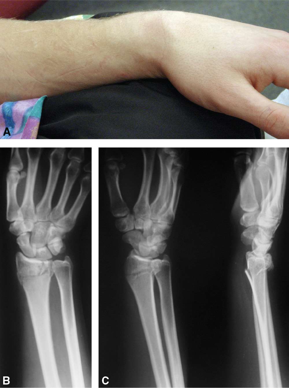
FIGURE 50.1 A: “Dinner-fork” deformity of Colles fracture. B, C: Colles fracture. (From Silverberg M. Colles’ and Smith’s fractures. In: Greenberg MI, ed. Greenberg’s Text-Atlas of Emergency Medicine. Philadelphia, PA: Lippincott Williams & Wilkins; 2005:483, with permission.)
Related posts:
Stay updated, free articles. Join our Telegram channel

Full access? Get Clinical Tree


