Chest pain (CP) is a common ED complaint. The management of these patients can be challenging, is of critical importance, and the clinician should systematically determine if CP is cardiac in origin or not.
PATHOPHYSIOLOGY
![]() CP can be visceral or somatic. Visceral pain originates from vessels or organs, enters the spinal cord at multiple levels, and is thus poorly described, using terms such as heaviness or aching. Somatic pain is dermatomal, from the parietal pleura or structures of the chest wall, and can be more precisely described.
CP can be visceral or somatic. Visceral pain originates from vessels or organs, enters the spinal cord at multiple levels, and is thus poorly described, using terms such as heaviness or aching. Somatic pain is dermatomal, from the parietal pleura or structures of the chest wall, and can be more precisely described.
![]() Ischemia is an imbalance of oxygen supply and demand.
Ischemia is an imbalance of oxygen supply and demand.
![]() Cardiac pain is visceral pain due to lack of oxygen to myocytes. Anaerobic metabolism ensues; chemical mediators are released and pain results.
Cardiac pain is visceral pain due to lack of oxygen to myocytes. Anaerobic metabolism ensues; chemical mediators are released and pain results.
CLINICAL FEATURES
![]() Typical ischemic CP is felt as tightness, squeezing, crushing, or pressure in the retrosternal/epigastric area. Radiation of pain to the jaw or arm is associated with a higher risk of ischemia; other symptoms also increase the chances of a cardiac etiology (see Table 19-1).
Typical ischemic CP is felt as tightness, squeezing, crushing, or pressure in the retrosternal/epigastric area. Radiation of pain to the jaw or arm is associated with a higher risk of ischemia; other symptoms also increase the chances of a cardiac etiology (see Table 19-1).
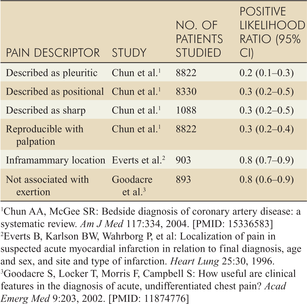
![]() Factors uncovered in the history and examination (such as pleuritc or positional pain) decrease the likelihood of ischemia or infarction (see Table 19-2), but do not rule out these more concerning conditions.
Factors uncovered in the history and examination (such as pleuritc or positional pain) decrease the likelihood of ischemia or infarction (see Table 19-2), but do not rule out these more concerning conditions.
![]() Symptoms lasting less than 2 minutes or continuously longer than 24 hours are less likely to be ischemic. Atypical presentations (anginal equivalents) are the rule and are more common in women, diabetics, the elderly, nonwhite minorities, and psychiatric patients.
Symptoms lasting less than 2 minutes or continuously longer than 24 hours are less likely to be ischemic. Atypical presentations (anginal equivalents) are the rule and are more common in women, diabetics, the elderly, nonwhite minorities, and psychiatric patients.
![]() Up to a third of patients with acute myocardial infarctions (AMIs) may not have CP. Patients may present with ischemic equivalents of shortness of breath (SOB), nausea, vomiting, shoulder or jaw pain, or palpitations.
Up to a third of patients with acute myocardial infarctions (AMIs) may not have CP. Patients may present with ischemic equivalents of shortness of breath (SOB), nausea, vomiting, shoulder or jaw pain, or palpitations.
![]() The character of the CP on presentation of the patient may aid in the differentiation of ischemic pain from other diagnostic possibilities.
The character of the CP on presentation of the patient may aid in the differentiation of ischemic pain from other diagnostic possibilities.
![]() Ischemic cardiac pain is typically worsened with exertion and relieved with rest.
Ischemic cardiac pain is typically worsened with exertion and relieved with rest.
DIAGNOSIS AND DIFFERENTIAL
![]() Diagnosis of cardiac CP is usually based on historical information.
Diagnosis of cardiac CP is usually based on historical information.
TABLE 19-1 Historical Factors That Increase the Likelihood of Acute Myocardial Infarction
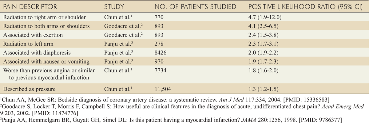
TABLE 19-2 Historical and Examination Factors That Decrease the Likelihood of Acute Myocardial Infarction
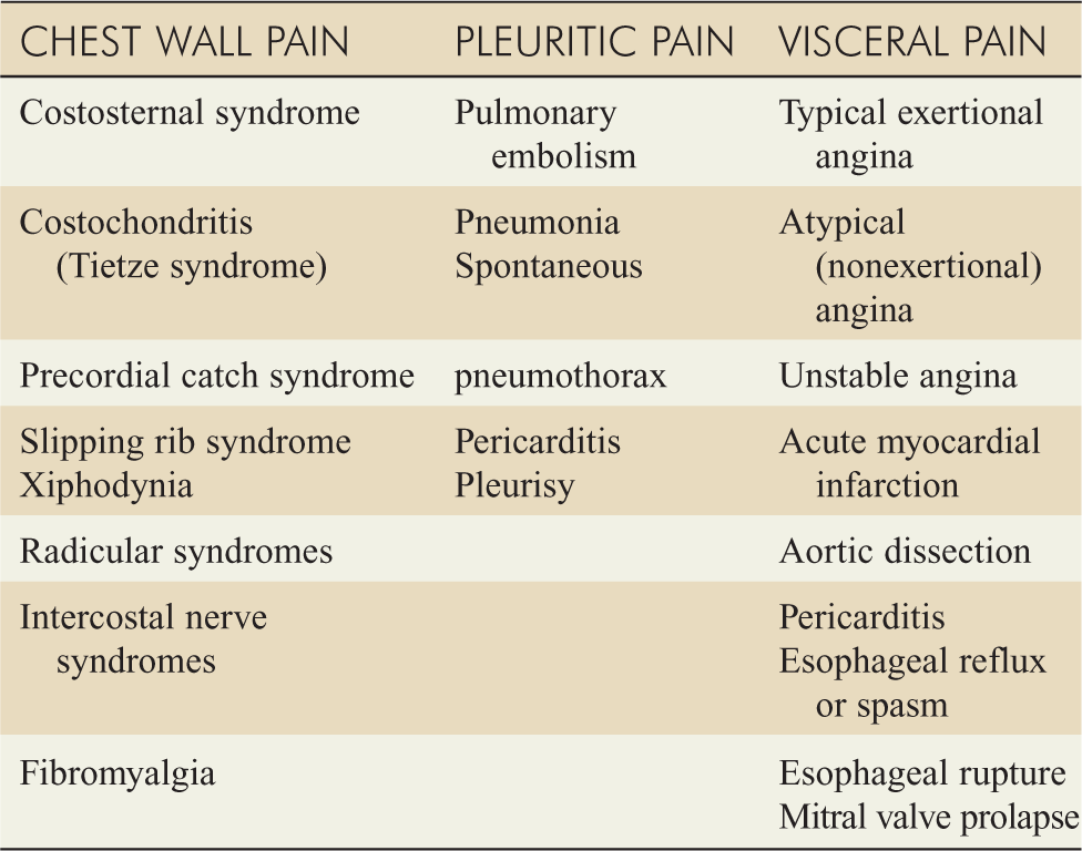
![]() There are many important causes of CP that may not be cardiac in nature (see Table 19-3).
There are many important causes of CP that may not be cardiac in nature (see Table 19-3).
![]() There are several life-threatening causes of CP which the emergency physician should consider for every CP patient (see Table 19-4).
There are several life-threatening causes of CP which the emergency physician should consider for every CP patient (see Table 19-4).
![]() The electrocardiogram (ECG) is the most important single test and should be obtained within 10 minutes of arrival to the ED. Only 50% of AMI patients will have a diagnostic ECG. Serial ECGs are imperative in the setting of continued CP.
The electrocardiogram (ECG) is the most important single test and should be obtained within 10 minutes of arrival to the ED. Only 50% of AMI patients will have a diagnostic ECG. Serial ECGs are imperative in the setting of continued CP.
TABLE 19-3 Important Causes of Acute Chest Pain
![]() Serum cardiac markers are useful for ischemia and infarction. Creatine Kinase (CK), specifically the MB fraction, measured over 24 hours historically has been considered the gold standard for AMI. However, CK-MB may be elevated by as many as 37 other varied conditions including extreme exercise, head trauma, and febrile disorders.
Serum cardiac markers are useful for ischemia and infarction. Creatine Kinase (CK), specifically the MB fraction, measured over 24 hours historically has been considered the gold standard for AMI. However, CK-MB may be elevated by as many as 37 other varied conditions including extreme exercise, head trauma, and febrile disorders.
![]() Troponin I and T are specific to cardiac muscle and have a high specificity and sensitivity for AMI. Troponins have thus replaced CK as the preferred cardiac marker. Troponins can also be elevated in noncardiac ischemic disorders (see Table 19-5).
Troponin I and T are specific to cardiac muscle and have a high specificity and sensitivity for AMI. Troponins have thus replaced CK as the preferred cardiac marker. Troponins can also be elevated in noncardiac ischemic disorders (see Table 19-5).
![]() Myoglobin rises predictably in AMI, but there is a high false-positive rate due to its presence in all muscle tissue.
Myoglobin rises predictably in AMI, but there is a high false-positive rate due to its presence in all muscle tissue.
TABLE 19-4 Life-Threatening Causes of Chest Pain: Classic Symptoms Compared*
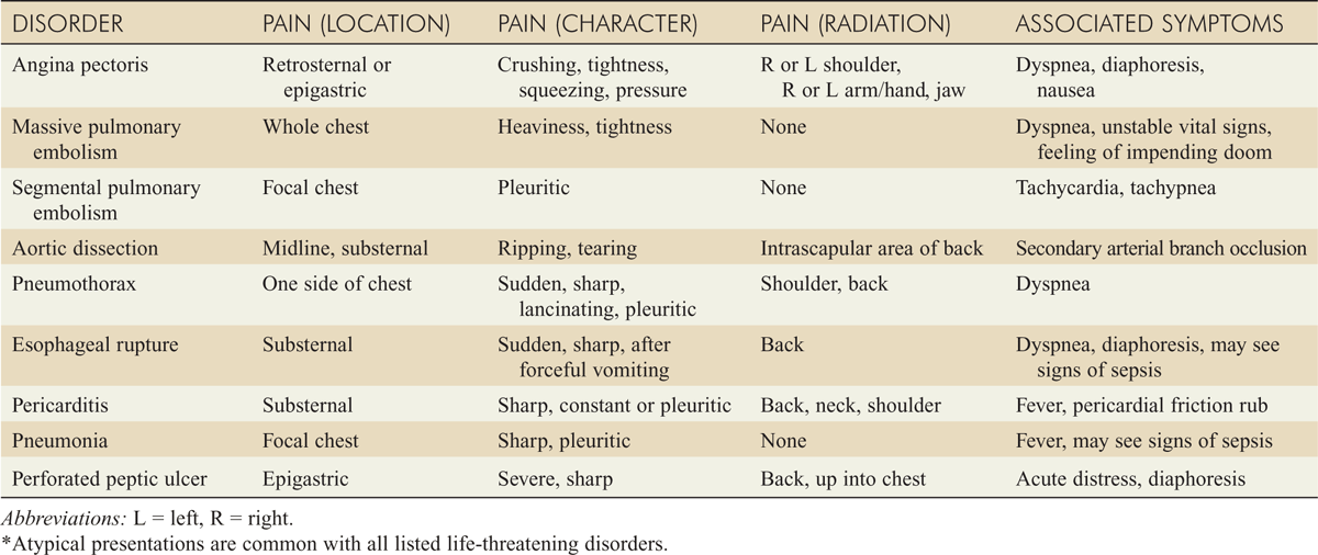
TABLE 19-5 Conditions Associated with Elevated Troponin Levels in the Absence of Ischemic Heart Disease
Cardiac contusion
Cardioinvasive procedures (surgery, ablation, pacing, stenting)
Acute or chronic congestive heart failure
Aortic dissection
Aortic valve disease
Hypertrophic cardiomyopathy
Arrhythmias (tachy- or brady-)
Apical ballooning syndrome
Rhabdomyolysis with cardiac injury
Severe pulmonary hypertension, including pulmonary embolism
Acute neurologic disease (eg, stroke, subarachnoid hemorrhage)
Myocardial infiltrative diseases (amyloid, sarcoid, hemochromatosis, scleroderma)
Inflammatory cardiac diseases (myocarditis, endocarditis, pericarditis)
Drug toxicity
Respiratory failure
Sepsis
Burns
Extreme exertion (eg, endurance athletes)
![]() The relationship between elevations of CK-MB, myoglobin, and troponin can be seen in Fig. 19-1.
The relationship between elevations of CK-MB, myoglobin, and troponin can be seen in Fig. 19-1.
![]() Various additional diagnostic tools can aid in the detection of cardiac CP. These are mentioned in detail in other chapters in this section but include echocardiography and stress testing (exercise, echo, and nuclear). These tests may aid in the diagnosis of cardiac CP; additionally, echocardiography can facilitate diagnosis of other conditions (eg, pericarditis, aortic dissection, hypertrophic cardiomyopathy) that can mimic ischemic disease.
Various additional diagnostic tools can aid in the detection of cardiac CP. These are mentioned in detail in other chapters in this section but include echocardiography and stress testing (exercise, echo, and nuclear). These tests may aid in the diagnosis of cardiac CP; additionally, echocardiography can facilitate diagnosis of other conditions (eg, pericarditis, aortic dissection, hypertrophic cardiomyopathy) that can mimic ischemic disease.
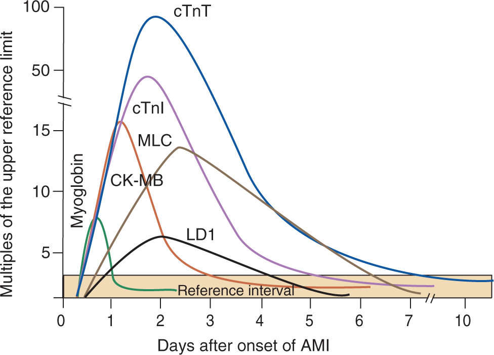
FIG. 19-1. Typical pattern of serum marker elevation after acute myocardial infarction (AMI). CK-MB = MB fraction of creatine kinase; cTnI = cardiac troponin I; cTnT = cardiac troponin T; LD1 = lactate dehydrogenase isoenzyme 1 ; MLC = myosin light chain.
![]() The use of chest x-ray usually provides limited information with cardiac CP, but chest x-ray can aid in the diagnosis of other serious conditions.
The use of chest x-ray usually provides limited information with cardiac CP, but chest x-ray can aid in the diagnosis of other serious conditions.
EMERGENCY DEPARTMENT CARE AND DISPOSITION
![]() Place the patient on a cardiac monitor, administer oxygen, and obtain IV access. Obtain an ECG. The initial focus should be on immediate life threats.
Place the patient on a cardiac monitor, administer oxygen, and obtain IV access. Obtain an ECG. The initial focus should be on immediate life threats.
![]() The patient should be treated aggressively if the clinical suspicion is high, even if the initial ECG is nondiagnostic of cardiac ischemia. Admit the patient to a high-acuity setting in the presence of an acute coronary syndrome.
The patient should be treated aggressively if the clinical suspicion is high, even if the initial ECG is nondiagnostic of cardiac ischemia. Admit the patient to a high-acuity setting in the presence of an acute coronary syndrome.
![]() The combination of ECG, clinical history, and cardiac markers should be used for both diagnostic purposes (ie, “is this pain cardiac?”) and for risk stratification that guides management and disposition decisions.
The combination of ECG, clinical history, and cardiac markers should be used for both diagnostic purposes (ie, “is this pain cardiac?”) and for risk stratification that guides management and disposition decisions.
Stay updated, free articles. Join our Telegram channel

Full access? Get Clinical Tree


