Cardiac Surgery

Chapter 88


Cardiac Surgery


The field of cardiac surgery has seen remarkable advances. Procedures that once required a large sternotomy incision and institution of cardiopulmonary bypass are now being done through small incisions in the chest with the heart still beating. Similarly, many procedures that historically required long periods of aortic and visceral ischemia in order to replace segments of aorta with prosthetic grafts are now being approached percutaneously and treated by the deployment of stent devices, obviating the need for prolonged malperfusion. Indeed, many of these patients do not require intensive care unit (ICU) attention postoperatively. Concomitant with these advances, however, is an aging population with a greater number of comorbid conditions, many of whom have previously undergone myriad interventional or open surgical procedures prior to presentation. It is therefore not surprising that the perioperative care of the cardiac surgical patient has become more complex, requiring the intensivist to have a thorough understanding not only of physiology but of the technical aspects of the devices and procedures. Although a comprehensive review of cardiac surgery is beyond the scope of this book, this chapter reviews the basic tenets of these complex operations and provides a pathophysiologic foundation upon which to approach the management of these patients in the ICU.


Effects of Cardiopulmonary Bypass


Understanding the cardiopulmonary bypass (CPB) circuit and the associated complications allows more effective and thoughtful care of these patients postoperatively. The CPB machine diverts venous blood through a membrane oxygenator, where oxygen is added and carbon dioxide is removed, and then returns this blood (now oxygenated and ventilated) to the arterial system at a physiologic pressure such that the perfusion to key organs remains adequate. Although simple in concept, the reality is that initiating CPB and exposing a patient’s entire cardiac output to foreign material, as well as converting organ perfusion from a state of pulsatile to continuous flow, create such enormous physiologic perturbations, at both at an organ and a cellular level, that the subsequent inflammatory cascade can be devastating if not managed appropriately in the perioperative period. Indeed, the majority of complications encountered after heart surgery can be attributed to the deleterious effects of CPB. To effectively manage the consequences of CPB, it is important to have a basic understanding of the bypass circuit, the potential complications that can arise while on CPB, and the ensuing inflammatory events that result from a patient’s blood interacting with artificial surfaces.


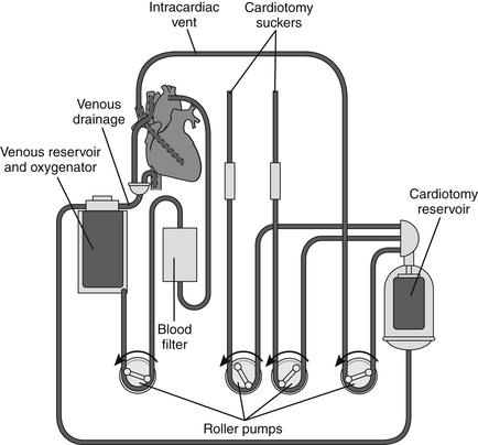
The basic CPB circuit (Figure 88.1) is composed of a large venous catheter (inflow), a reservoir, a centrifugal or roller pump, an oxygenator, a heat exchanger, a debubbler, and an arterial catheter (outflow). Inflow of venous blood to the reservoir occurs primarily by gravity. Depending on the procedure, these venous catheters can be placed either centrally (e.g., right atrium, inferior vena cava, superior vena cava) or peripherally (e.g., femoral vein, internal jugular vein). Blood loss in the operative field is collected by pump suckers and also returned to the venous reservoir. Reservoir blood is then pumped through the oxygenator and heat exchanger. In turn, the warmed and oxygenated blood is infused into the arterial system via a cannula placed either centrally (e.g., ascending aorta, aortic arch) or peripherally (e.g., femoral or axillary artery).


Essential to this entire process is the need for profound systemic anticoagulation to prevent thrombin formation in the pump. This is usually attained by the administration of large amounts of unfractionated heparin and can be followed by frequent measurements of activated clotting times (ACTs). Typically, heparin is administered prior to aortic or venous cannulation, and the ACT is verified to be > 350 to 400 seconds. Heparin titration protocols have been developed based on individual dose response curves to ensure the patient is not under- or overdosed with heparin.


Exposure of blood to the CPB circuit results in a massive systemic inflammatory response syndrome (SIRS), which, if left unchecked, ultimately manifests as end organ dysfunction in the early postoperative period. These complications are often magnified in patients who sustain prolonged bypass times, patients with preexisting organ dysfunction, or patients who sustain extremes in blood pressure during the operation.


Complement activation induced by exposure of plasma to the foreign CPB circuit causes a generalized inflammatory reaction characterized by the release of multiple cytokines, lymphokines, and proteases. Furthermore, this cascade of events augments platelet activation, coagulation, and fibrinolysis. The resulting coagulation contributes to ischemia-reperfusion injury in various end organs. Platelet disruption occurs as a result of direct contact with the circuit’s inner surface, especially at the level of the oxygenator. Platelet dysfunction also occurs as a consequence of complement-induced opsonization in addition to the generalized inflammatory state. Ultimately, the fibrinolytic pathway is activated by CPB increasing levels of tissue plasminogen activator (tPA) in response to endothelial damage, platelet activation, and complement activation. Hemodilution and consumption of these clotting factors contribute to a bleeding diathesis postoperatively. Red blood cell disruption, which manifests as hemoglobinuria, correlates directly with the intensity of pump sucker use as well as with the total duration of CPB. White blood cell activation and subsequent sequestration in the lungs can occur as a result of CPB. When combined with complement activation and high levels of circulating cytokines, this can lead to pulmonary edema and injury and, rarely, postoperative acute respiratory distress syndrome (ARDS).


Myocardial Protection


An essential component to most cardiac surgical procedures is ensuring viability of the heart while the aortic cross-clamp is in place (the period of cardiac ischemia). When CPB is first initiated, blood flow and oxygenation to the heart persist (via flow through coronary arteries). However, many operations on the heart require a bloodless field and cessation of the heart’s contractions. In order to exclude the heart from the bypassed blood flow, a clamp is placed on the ascending aorta, proximal to the arterial blood outflow from CPB, inhibiting blood from perfusing the myocardium. Although this ischemic period is necessary, a number of methods aimed at reducing myocardial oxygen consumption, restoring a modest amount of blood flow to the heart, and ensuring the heart remains empty optimize the conditions during the myocardial ischemic period, making it easier to come off CPB after cardiac reperfusion.


Decreasing myocardial oxygen consumption minimizes the need to constantly supply blood flow to the heart. Delivery of a high potassium concentration solution (cardioplegia) into the aortic root, proximal to the aortic cross clamp, and subsequently into the coronary arteries (assuming the aortic valve is competent) will arrest the heart in diastole, immediately decreasing oxygen consumption by at least 90%. Another key aspect of myocardial protection is ensuring that the heart remains empty throughout the procedure and does not fill with blood, causing distention of the cavities and increased myocardial oxygen consumption. This is attained by placement of catheters (“vents”), which continuously divert any intracardiac blood back into the CPB circuit.


Finally, both myocardial and systemic hypothermia are induced intraoperatively to help minimize oxygen consumption of all organs. Typically, cardioplegia is delivered to the heart at 9.0° to 15.0° C and the body is cooled to 32.0° C during CPB. This is achieved by cooling the blood externally while it is in the CPB circuit.


Evaluation of Cardiac Function in the Intensive Care Unit


In general, all patients are routinely admitted to the ICU after heart surgery. Many of these patients arrive to the ICU intubated with invasive hemodynamic monitoring and are receiving a variety of vasoactive infusions. It is essential in the early postoperative period to monitor recovery of heart function and recovery from anesthesia, to accurately quantify bleeding from chest tubes, and to evaluate end organ function.


Invasive Hemodynamic Monitoring


Various forms of invasive hemodynamic monitoring are used to guide postoperative recovery of heart function, fluid management, and overall management in the ICU setting (Chapter 7). Continuous blood pressure is monitored via a radial or femoral arterial catheter. Many patients also have a pulmonary arterial catheter (PAC) in place, which can be helpful in assessing both right and left heart function. Importantly, in patients with a PAC, great caution should be taken if a pulmonary artery wedge pressure (PAWP) is deemed necessary, as many of these patients have multiple factors, including pulmonary hypertension and an acquired bleeding diathesis, that put them at high risk for balloon-induced rupture of the pulmonary artery, a usually fatal complication.

< div class='tao-gold-member'>

Stay updated, free articles. Join our Telegram channel

Jul 7, 2016 | Posted by in CRITICAL CARE | Comments Off on Cardiac Surgery

Full access? Get Clinical Tree

Get Clinical Tree app for offline access