 Hemodynamic instability (presyncope, angina, altered mentation, pulmonary edema) secondary to:
 Bradydysrhythmias:
 Sinus bradycardia
 Sinus node dysfunction
 Atrioventricular (AV) node conduction blocks (second and third degree)
 Acute myocardial infarction with bifascicular block, alternating bundle branch block, new left bundle branch block (LBBB), escape rhythm <40 bpm
 Malfunctioning permanent pacemaker
 Electrolyte/metabolic disturbances (i.e., hyperkalemia) if medical therapies fail or are unavailable
 Post–cardiac surgery (i.e., valve replacement)
 Thoracic trauma (i.e., cardiac contusion)
 Other (i.e., lyme carditis, endocarditis)
 Tachydysrhythmias (overdrive pacing, typically transvenous only):
 Supraventricular tachycardia (SVT)
 Ventricular tachycardia (VT)
 Transvenous Pacing—same as above, but also including:
 Failure or nontolerance of transcutaneous pacing
 Bridge to permanent pacemaker placement
 High risk of progression to complete heart block
 Overdrive pacing
CONTRAINDICATIONS
 Absolute
 Asymptomatic, stable rhythms (i.e., first-degree AV block)
 Prosthetic tricuspid valve (transvenous only)
 Relative
 Severe hypothermia (may be physiologic bradycardia; can induce fibrillation)
 Brady-asystolic arrest >20 minutes
 Drug-induced dysrhythmias (although can be utilized as last resort if antidote fails)
TRANSCUTANEOUS PACING PROCEDURE
 Landmarks
Pacer pads should preferentially be placed over the precordium anteriorly and in the interscapular paraspinous region posteriorly. Anterolateral pad placement is also acceptable.
 Supplies
 Cardiac monitor with pacemaker capabilities (if electrocardiogram [ECG] monitor is not part of the pacer unit, a separate monitor and adaptor will be required)
 Adhesive pacing pads
 ECG electrodes
 Safety razor
 Technique
 Preparation
 In conscious patients, reassurance and explanation of the procedure, including expectations for discomfort, are extremely important
 Remove excess hair if time permits
 Continuous cardiac and pulse oximetry monitoring, intravenous access, and bedside capability for resuscitation, including airway management, defibrillation, and arrhythmia treatment, should be at the bedside before initiation
 Pacer/Electrode Placement
 Pads are placed as shown in FIGURE 9.1
 Anterior chest pacing pad (negative charge electrode) is placed over the point of maximal impulse
 If access to the posterior chest wall is limited or difficult, posterior pacer pad may also be placed in cardiac apex/base position (identical to electrical cardioversion placement)
 ECG electrodes should be placed in limb lead positions for monitoring
 Pacing
 Identify pacemaker mode on equipment and turn to “on”
 Set heart rate to 70 bpm
 Place and maintain one hand in pulse-check position (radial, femoral, or carotid) or observe noninvasive or invasive blood pressure response
 In bradyasystole and unconscious patients, set current to 150 to 200 mA and lower in 10-mA decrements; set current at the lowest level that will consistently achieve mechanical capture
 In stable and conscious patients, set current to 10 mA and raise in 10-mA increments until mechanical capture is achieved
 Observe cardiac monitor for pacemaker spikes and electrical capture—“electrical capture” refers to narrow pacemaker spikes followed by typically wide ventricular complexes
 Monitor constantly for “mechanical capture”—a palpable arterial pulse induced by pacemaker discharges or perfusing blood pressure by noninvasive or invasive blood pressure monitoring
 Titrate sedation/analgesia/anxiolysis to allow for tolerance of ongoing pacing
 Failure to achieve mechanical capture should prompt immediate preparation for transvenous pacer placement
 Complications
 Unrecognized ventricular fibrillation
 Local discomfort
 Cutaneous injury
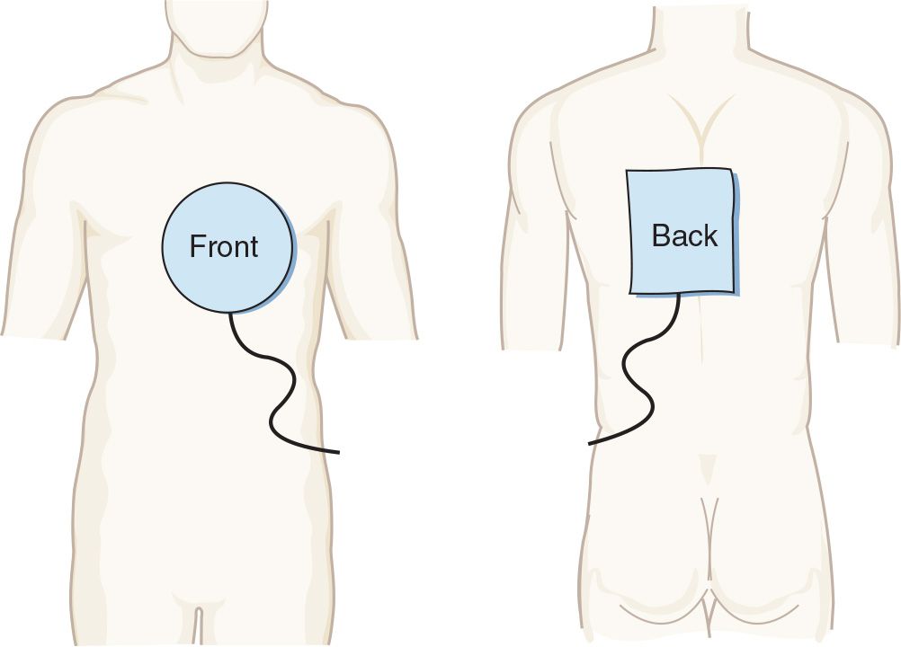
FIGURE 9.1 Proper placement of transcutaneous pacing electrodes. (From Morton PG, Fontaine DK. Critical Care Nursing. 10th ed. Philadelphia, PA: Wolters Kluwer Health; 2012.)
SAFETY/QUALITY TIPS
 Procedural
 The most common causes of failure to capture in transcutaneous pacing are improper electrode placement or large patient size
 For many patients, adequate amperage will not be possible without aggressive sedation/analgesia
 Cognitive
 Compared to transvenous pacing, transcutaneous pacing is painful and ineffective. Transcutaneous pacing should be thought of as a brief bridge to transvenous pacing or correction of the underlying disorder.
 Electrical capture is not mechanical capture, and mechanical capture is what counts. Once electrical capture occurs, mechanical capture must be immediately verified using pulses, invasive arterial pressure monitoring, ultrasound, or (most conveniently) pulse oximetry.
 Be careful not to mistake ventricular fibrillation or tachycardia for a paced rhythm
Related posts:



Stay updated, free articles. Join our Telegram channel

Full access? Get Clinical Tree


