and Richard A. Jaffe2
(1)
David Geffen School of Medicine at UCLA, Los Angeles, California, USA
(2)
Stanford University School of Medicine, Stanford, California, USA
Keywords
BronchospasmWheezingAirway obstructionLung volumeAtelectasisIntroduction
There is a well-known adage, which says “all that wheezes is not asthma”. It is equally true that “all that wheezes during anesthesia is not bronchospasm”. It is a widely held view among anesthesia providers that bronchospasm is relatively common during anesthesia. If a large audience of anesthesia providers is asked “How many of you have experienced a case of bronchospasm?” virtually everyone’s hands will go up. If they are asked how many have seen multiple cases of bronchospasm, most will raise their hands again. Most anesthesia providers believe that patients with a history of asthma or other bronchospastic disorder have persistent airway hyperreactivity, which may result in intense bronchospasm during anesthesia [1, 2]. We do not share these views. One of the authors has never seen a documented case of bronchospasm in over 50+ years of clinical practice. He has seen many cases where it was thought that bronchospasm was occurring, but on careful examination it proved to be the wrong diagnosis. In our opinion, bronchospasm is a relatively rare cause of wheezing during anesthesia, and because of this fact, it is one of the most misunderstood, misdiagnosed, and mistreated conditions in clinical anesthesia.
When managing a patient under general anesthesia who develops wheezing and requires increased airway pressures to maintain adequate ventilation, the first diagnosis the anesthesia provider should consider is partial airway obstruction . Kinking of the tracheal tube or partial occlusion of the tube with secretions or blood may produce wheezing-like sounds during ventilation along with elevation of peak airway pressure. The first step in diagnosis and treatment is to deflate the tracheal tube cuff, suction the tracheal tube, perhaps rotate the tube 45° and establish that it is functioning properly. Once that is done the anesthesia provider should direct attention to passive or active bronchoconstriction.
Both active and passive forces regulate airway size . A decrease in airway size may be caused by either active or passive forces (Table 7.1).
Table 7.1
Increased airway resistance during anesthesia forces
Forces | ||
|---|---|---|
Passive | Active | |
Incidence | Common | Rare |
Etiology | Loss of lung volume | Histamine release |
Stimulation airway receptors | ||
Decreased breath sounds | Yes | Yes |
Wheezing present | Yes | Yes |
Decreased lung compliance | Yes | Yes |
Increased peak airway pressure | Yes | Yes |
Abdominal wall muscle tone | Increased | Unchanged |
Treatment | Increase lung volume | Albuterol aerosol |
Deepen anesthesia | Deepen anesthesia | |
Epinephrine if anaphylaxis | ||
Bronchodilators helpful | No | Yes |
NMB drugs helpful | Yes | No |
There are two active forces which either directly or indirectly cause an increase in smooth muscle tone of the airways to produce constriction. One of the active forces is the autonomic nervous system. The airways are richly innervated by the parasympathetic nervous system , which plays a major role in the constriction of the airways. It is believed that stimulation of irritant receptors in the large airways, for example by the insertion of a tracheal tube, causes a release of acetylcholine from parasympathetic nerves that stimulates M2 and M3 muscarinic receptors in airway smooth muscle causing bronchoconstriction [2, 3]. However, this bronchoconstrictive reflex is easily interrupted by anesthetics. For example, the afferent limb is readily blocked by the topical application of local anesthetics in the airway. As well, the efferent limb is blocked by atropine, and the entire reflex is attenuated by general anesthesia or systemically-administered local anesthetics. Hickey et al. showed in dogs that 0.5 MAC halothane blocked vagal bronchoconstriction induced by electrical stimulation of the vagus nerve [4]. Sevoflurane, isoflurane, and halothane have all been shown to be effective bronchodilators at low concentrations.
The second active force regulating airway size is chemical. The most important chemical mediator is histamine , which is contained in mast cells in the airways. Histamine released by anesthetic drugs acts on H1 receptors in the airway to cause bronchoconstriction of airway smooth muscle. The most dramatic and very rare form of this is anaphylaxis, which within 5 min of administration of the offending anesthetic drug is manifest by wheezing (bronchospasm), decrease in pulmonary compliance, marked decrease in blood pressure, and bradycardia . If not treated promptly with epinephrine, a cardiac arrest may occur. The neuromuscular blocking drugs rocuronium and vecuronium are the most common anesthetic drugs causing anaphylaxis.
Both alpha and beta2 receptors are present in airway smooth muscle. Stimulation of alpha receptors causes airway constriction, but the reflex is weak and probably not important in the regulation of airway size. Stimulation of beta2 receptors, for example by circulating epinephrine, activates adenyl cyclase to increase the concentration of cyclic AMP, which in turn causes relaxation of airway smooth muscle [2]. Beta adrenergic blocking drugs (e.g. propranolol) will block this reflex and this may be the mechanism for bronchoconstriction observed in some patients given these drugs. Whether inhalation anesthetics cause bronchodilatation by stimulation of beta2 receptors is not known, but there is evidence that ketamine may cause bronchodilatation by this mechanism. Instead it is generally accepted that inhalation anesthetics act directly on airway smooth muscle to produce bronchodilatation. Maximum dilatation will occur at 0.5 MAC or less.
Normal patients and even most patients who have a history of asthma or other bronchospastic disorders have normal airway resistance when the disease is in remission. At normal lung volumes, airways are at or near maximal dilatation and measurements of airway resistance are in the normal range of 1 cmH2O/L/s. or less. Because of this, the administration of bronchodilator drugs does not increase airway size or decrease airway resistance.
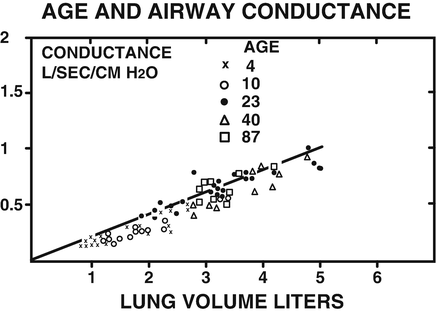
What is not widely recognized or appreciated is that the single most important determinant of airway size is lung volume. As lung volume increases during spontaneous ventilation, the increasing negative intrathoracic pressure and expanding alveoli apply radial traction on all of the airways and they enlarge. During mechanical ventilation, positive airway pressure causes airway expansion. With this change, airway resistance decreases and airway conductance (the reciprocal of resistance) increases [5]. Even the large airways such as the trachea and major bronchi enlarge. The reverse occurs as lung volume decreases. The decrease in intrathoracic pressure and loss of radial traction on the airways cause them to narrow. The greater the loss in lung volume, the more constricted the airways become. Briscoe and Dubois demonstrated in 26 patients of varying size and age that there was a close correlation between lung volume and airway resistance and airway conductance (Fig. 7.1) [6]. Tammeling and Sluiter demonstrated that even the tracheal diameter is affected by lung volume (Fig. 7.2) [7]. In 19 healthy men, intrathoracic tracheal diameter ranged from 80 to 100 % of maximum during maximal inspiration, and ranged from 20 to 60 % of maximum during maximal expiration. Clearly with maximal exhalation tracheal diameter becomes markedly narrowed.