| Sick sinus syndrome |
| Infiltrative diseases (e.g., sarcoidosis) |
| Collagen vascular diseases (e.g., scleroderma) |
| Atrioventricular conduction block |
| Acute or chronic ischemic heart disease |
| Increased vagal tone (e.g., Valsalva maneuver) |
| Drugs (e.g., digoxin, beta-blockers) |
| Obstructive sleep apnea |
| Hypothyroidism |
| Hypothermia |
| Vector-borne illness (e.g., Lyme disease) |
| Increased intracranial pressure (i.e., Cushing reflex) |
| Electrolyte disturbances (e.g., hypo/hyperkalemia) |
Presentation
Classic presentation
- Although the symptoms are nonspecific, many will be related to inadequate cardiac output secondary to the low heart rate:
- Orthostatic hypotension
- Dizziness
- Syncope
- Generalized fatigue or malaise
- Chest pain
- Shortness of breath
- Orthostatic hypotension
Critical presentation
- Sinus bradycardia occurs in 15–20% of patients with acute myocardial infarction secondary to ischemia of the SA node.
- Syncope may result from primary dysrhythmia or from reduced cardiac output.
- Hemodynamic instability:
- Similarly to patients with rapid heart rates, instability is defined as the evidence of hemodynamic compromise with a discernible pulse.
- Patients will present with hypotension, altered mental status, ischemic chest pain, or respiratory distress from congestive heart failure and pulmonary edema.
- Patients who do not have a palpable pulse are in cardiac arrest and are treated according to ACLS guidelines.
- Similarly to patients with rapid heart rates, instability is defined as the evidence of hemodynamic compromise with a discernible pulse.
Diagnosis and evaluation
- A 12-lead electrocardiogram (ECG) is essential for the diagnosis of bradycardia and to differentiate between the different types of bradyarrhythmias.
- History should focus particularly on symptoms of ischemic heart disease, and on medications such as nodal blockers.
- Some laboratory values may be helpful, depending on the suspected etiology:
- Troponin to assess for myocardial injury
- Electrolytes to rule out hyperkalemia from changes in renal function
- Digoxin level if appropriate
- Thyroid-stimulating hormone (TSH).
- Troponin to assess for myocardial injury
- A rectal temperature should be taken to rule out hypothermia as a possible etiology.
- Once the ECG is performed, further information about the location and severity of the conduction delay can be obtained.
- SA node dysfunction:
- Irregular or absent sinus node activity.
- Most common reason for pacemakers in the United States.
- “Sick sinus syndrome”:
- Caused by a worsening fibrosis of the SA node or diminished blood flow to the SA nodal artery.
- Most often seen in patients older than 70 years old.
- Characteristics include inappropriate bradycardia, alternating bradycardia and tachyarrhythmias, and/or sinus pauses or sinus arrest.
- Caused by a worsening fibrosis of the SA node or diminished blood flow to the SA nodal artery.
- Irregular or absent sinus node activity.
- SA node dysfunction:
- AV node dysfunction:
- AV conduction blocks can be either transient or permanent and can result from numerous etiologies.
- The most common cause of AV nodal dysfunction is ischemic disease (40%), though other etiologies are possible:
- Idiopathic fibrosis of the conduction system
- Bacterial endocarditis
- Vector-borne disease
- Medication overdose
- Idiopathic fibrosis of the conduction system
- AV conduction blocks can be either transient or permanent and can result from numerous etiologies.
- AV node dysfunction can be classified as first-degree, second-degree (types I and II) or third-degree (i.e., complete heart block).
- First-degree AV block:
- Defined as a PR interval of greater than 200 milliseconds.
- Each P is associated with a QRS and both the P-P and R-R intervals remain constant.
- It is generally considered to be benign.
- Defined as a PR interval of greater than 200 milliseconds.
- First-degree AV block:
- Second-degree AV block: Mobitz type I (aka: Wenckebach (see Figure 22.1):
- Defined as a rhythm with an increasingly prolonged PR interval that will eventually lead to a dropped beat.
- This pattern can happen intermittently or persistently, and in a variety of groupings.
- Like first-degree AV blocks it is generally considered to be benign and does not require intervention.
- Defined as a rhythm with an increasingly prolonged PR interval that will eventually lead to a dropped beat.
- Second-degree AV block: Mobitz type II (see Figure 22.2):
- Defined as a consistent PR interval with intermittent failure of conduction through the AV node resulting in dropped beats.
- Can be seen in grouped beats as type I, but can also have varying degrees of block.
- Frequently progresses to complete heart block (third-degree heart block).
- Due to the potential for progression of the blockade, these patients require admission to the hospital and evaluation for a pacemaker.
- Defined as a consistent PR interval with intermittent failure of conduction through the AV node resulting in dropped beats.
- Third-degree AV block (complete heart block) (see Figure 22.3):
- Defined as a complete disassociation between atrial and ventricular activity.
- No relationship exists between P-waves and the QRS complexes.
- Ventricular escape beats can originate anywhere from the AV node (narrow complex) to the Purkinje system (wide QRS complex).
- A permanent pacemaker is indicated in the setting of this rhythm.
- Defined as a complete disassociation between atrial and ventricular activity.
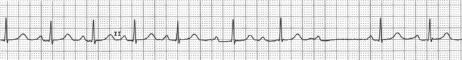
Figure 22.1. Second-degree AV block: Mobitz type I (Wenckebach).
Related posts:


Stay updated, free articles. Join our Telegram channel

Full access? Get Clinical Tree


