![]() Clinical suspicion of cholecystitis or biliary colic:
Clinical suspicion of cholecystitis or biliary colic:
![]() Abdominal pain, particularly right upper quadrant (RUQ), epigastric
Abdominal pain, particularly right upper quadrant (RUQ), epigastric
![]() Right flank pain
Right flank pain
![]() Right shoulder pain
Right shoulder pain
![]() Nausea/vomiting
Nausea/vomiting
![]() Sepsis without source
Sepsis without source
CONTRAINDICATIONS
![]() None: No contrast or radiation
None: No contrast or radiation
RISKS/CONSENT ISSUES
![]() Allergy to the ultrasonography gel
Allergy to the ultrasonography gel
LANDMARKS
![]() RUQ of abdomen at expected location of gallbladder (GB)
RUQ of abdomen at expected location of gallbladder (GB)
TECHNIQUE
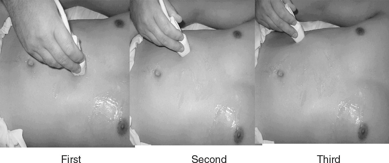
![]() Using a standard 3.5- to 5.0-MHz probe, scan the RUQ of the abdomen in the longitudinal plane under the costal margin using the liver as an acoustic window (FIGURE 34.1)
Using a standard 3.5- to 5.0-MHz probe, scan the RUQ of the abdomen in the longitudinal plane under the costal margin using the liver as an acoustic window (FIGURE 34.1)
![]() If the GB is not readily identified:
If the GB is not readily identified:
![]() Ask the patient to take slow deep breaths because the GB moves significantly with respiration
Ask the patient to take slow deep breaths because the GB moves significantly with respiration
![]() Have the patient move to the left lateral decubitus position
Have the patient move to the left lateral decubitus position
![]() Place the probe in the intercostal space to avoid bowel gas and rib shadows
Place the probe in the intercostal space to avoid bowel gas and rib shadows
![]() Once the GB is found, confirm by identifying associated structures:
Once the GB is found, confirm by identifying associated structures:
![]() Echogenic gallstones in the lumen casting echolucent shadows (FIGURE 34.2)
Echogenic gallstones in the lumen casting echolucent shadows (FIGURE 34.2)
![]() Main lobar fissure of the liver points toward the GB neck and also connects the portal vein
Main lobar fissure of the liver points toward the GB neck and also connects the portal vein
![]() Common bile duct (CBD) usually runs between the GB and the portal vein (FIGURE 34.3)
Common bile duct (CBD) usually runs between the GB and the portal vein (FIGURE 34.3)
![]() Identify signs of cholecystitis (TABLE 34.1)
Identify signs of cholecystitis (TABLE 34.1)
Related posts:
Stay updated, free articles. Join our Telegram channel

Full access? Get Clinical Tree


