![]() To assess for the absence of lung sliding, suggestive of a pneumothorax, in the following conditions:
To assess for the absence of lung sliding, suggestive of a pneumothorax, in the following conditions:
![]() Blunt thoracoabdominal trauma
Blunt thoracoabdominal trauma
![]() Penetrating thoracoabdominal trauma
Penetrating thoracoabdominal trauma
![]() Unexplained hypotension
Unexplained hypotension
![]() To assess for the presence of pleural fluid, suggestive of a hemothorax, in the following conditions:
To assess for the presence of pleural fluid, suggestive of a hemothorax, in the following conditions:
![]() Blunt thoracoabdominal trauma
Blunt thoracoabdominal trauma
![]() Penetrating thoracoabdominal trauma
Penetrating thoracoabdominal trauma
![]() Unexplained hypotension
Unexplained hypotension
CONTRAINDICATIONS
![]() If the EFAST examination delays a patient’s transport to the operating room
If the EFAST examination delays a patient’s transport to the operating room
![]() Theoretical allergy to the ultrasound gel
Theoretical allergy to the ultrasound gel
ADVANTAGES
![]() Noninvasive
Noninvasive
![]() No sedation required
No sedation required
![]() Performed at the bedside amidst simultaneous resuscitative efforts
Performed at the bedside amidst simultaneous resuscitative efforts
![]() Does not require transportation to the radiology suite
Does not require transportation to the radiology suite
![]() Serial examinations may be performed with changes in symptoms or hemodynamics
Serial examinations may be performed with changes in symptoms or hemodynamics
LANDMARKS
![]() Anterior Thorax
Anterior Thorax
![]() The apical midclavicular line in the sagittal plane; transducer marker positioned cephalad
The apical midclavicular line in the sagittal plane; transducer marker positioned cephalad
![]() Lateral Thorax
Lateral Thorax
![]() The lateral thorax in the axillary region; transducer marker positioned obliquely and cephalad
The lateral thorax in the axillary region; transducer marker positioned obliquely and cephalad
![]() Pleural (Right: Hepatorenal and Left: Splenorenal)
Pleural (Right: Hepatorenal and Left: Splenorenal)
![]() Transducer placed in the axillary line in the coronal plane at the level of 8th and 11th ribs; anterior axillary line on the right and posterior axillary line on the left, with the diaphragm as a landmark; transducer marker positioned toward the axilla.
Transducer placed in the axillary line in the coronal plane at the level of 8th and 11th ribs; anterior axillary line on the right and posterior axillary line on the left, with the diaphragm as a landmark; transducer marker positioned toward the axilla.
TECHNIQUE
![]() The EFAST standard views in addition to the basic FAST examination:
The EFAST standard views in addition to the basic FAST examination:
![]() Bilateral anterior thorax
Bilateral anterior thorax
![]() Bilateral lateral thorax
Bilateral lateral thorax
![]() Bilateral pleural spaces
Bilateral pleural spaces
![]() Anterior Thorax
Anterior Thorax
![]() Place the transducer in the second or third intercostal space in the midclavicular line
Place the transducer in the second or third intercostal space in the midclavicular line
![]() The indicator should be cephalad
The indicator should be cephalad
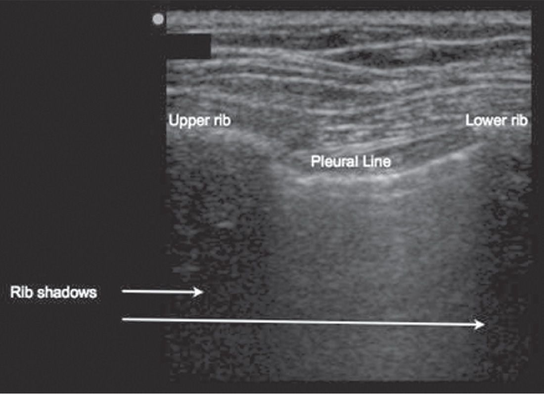
![]() Identify the bat sign: The upper rib–pleural line–lower rib profile (FIGURE 19.1)
Identify the bat sign: The upper rib–pleural line–lower rib profile (FIGURE 19.1)
![]() Normal lung findings
Normal lung findings
![]() B-mode: Visible sliding (shimmering or twinkling) at the level of the pleura
B-mode: Visible sliding (shimmering or twinkling) at the level of the pleura
![]() B-mode: Comet tails—vertical reverberation artifacts arising from the pleural line (FIGURE 19.2)
B-mode: Comet tails—vertical reverberation artifacts arising from the pleural line (FIGURE 19.2)
![]() M-mode: Seashore sign (FIGURE 19.3)
M-mode: Seashore sign (FIGURE 19.3)
![]() Pneumothorax
Pneumothorax
![]() B-mode: Loss of pleural sliding, as there is loss of contact between the visceral and the parietal pleura
B-mode: Loss of pleural sliding, as there is loss of contact between the visceral and the parietal pleura
![]() B-mode: Absence of comet tails
B-mode: Absence of comet tails
![]() M-mode: Stratosphere sign or bar-code sign (FIGURE 19.4)
M-mode: Stratosphere sign or bar-code sign (FIGURE 19.4)
![]() Lung point: Transition between collapsed and normally expanded lung; 100% specific for pneumothorax when identifiable
Lung point: Transition between collapsed and normally expanded lung; 100% specific for pneumothorax when identifiable
![]() TABLE 19.1 compares the signs suggestive of normal lung with those of pneumothorax
TABLE 19.1 compares the signs suggestive of normal lung with those of pneumothorax
Related posts:
Stay updated, free articles. Join our Telegram channel

Full access? Get Clinical Tree


