![]() Diagnostic
Diagnostic
![]() Evacuate abnormal collections of fluid from the joint space for synovial fluid analysis of the following suspected conditions:
Evacuate abnormal collections of fluid from the joint space for synovial fluid analysis of the following suspected conditions:
![]() Septic arthritis
Septic arthritis
![]() Crystal arthropathy
Crystal arthropathy
![]() Hemarthrosis
Hemarthrosis
![]() Inflammatory process
Inflammatory process
![]() Diagnose occult fracture or ligamentous injury
Diagnose occult fracture or ligamentous injury
![]() Inject sterile saline to test for joint capsule integrity when overlying laceration potentially extends into joint space
Inject sterile saline to test for joint capsule integrity when overlying laceration potentially extends into joint space
![]() Therapeutic
Therapeutic
![]() Drain effusion to decrease/relieve pressure in the joint to provide pain relief
Drain effusion to decrease/relieve pressure in the joint to provide pain relief
![]() Instill medication for treatment and pain relief
Instill medication for treatment and pain relief
CONTRAINDICATIONS
![]() Absolute Contraindications
Absolute Contraindications
![]() Abscess/cellulitis in the tissues overlying the site to be punctured (often infectious arthritis can mimic an overlying soft-tissue infection)
Abscess/cellulitis in the tissues overlying the site to be punctured (often infectious arthritis can mimic an overlying soft-tissue infection)
![]() Relative Contraindications
Relative Contraindications
![]() Bleeding diatheses or anticoagulant therapy
Bleeding diatheses or anticoagulant therapy
![]() Known bacteremia
Known bacteremia
![]() Prosthetic joint
Prosthetic joint
RISKS/CONSENT ISSUES
![]() Potential for introducing infection (sterile technique must be utilized)
Potential for introducing infection (sterile technique must be utilized)
![]() Procedure can cause pain and discomfort (local anesthesia will be given)
Procedure can cause pain and discomfort (local anesthesia will be given)
![]() Needle puncture can cause localized bleeding
Needle puncture can cause localized bleeding
![]() Reaccumulation of fluid may occur
Reaccumulation of fluid may occur
![]() Risk of injuring articular cartilage with needle tip
Risk of injuring articular cartilage with needle tip
![]() Potential for tendon and nerve damage if a medication is incorrectly instilled
Potential for tendon and nerve damage if a medication is incorrectly instilled
![]() General Basic Steps
General Basic Steps
![]() Position patient
Position patient
![]() Analgesia
Analgesia
![]() Aspiration
Aspiration
![]() Fluid analysis
Fluid analysis
LANDMARKS
![]() Two approaches are available (FIGURE 57.1):
Two approaches are available (FIGURE 57.1):
![]() Medial approach (most common):
Medial approach (most common):
![]() Identify the malleolar sulcus which allows a portal to the tibiotalar joint space. It is a small depression that is bordered by the medial malleolus medially and the anterior tibial tendon laterally.
Identify the malleolar sulcus which allows a portal to the tibiotalar joint space. It is a small depression that is bordered by the medial malleolus medially and the anterior tibial tendon laterally.
![]() Be wary of the saphenous vein and nerve, which lie laterally to medial malleolus
Be wary of the saphenous vein and nerve, which lie laterally to medial malleolus
![]() Lateral approach: The subtalar joint space lies approximately ½ inch proximal and medial to the distal tip of the lateral malleolus
Lateral approach: The subtalar joint space lies approximately ½ inch proximal and medial to the distal tip of the lateral malleolus
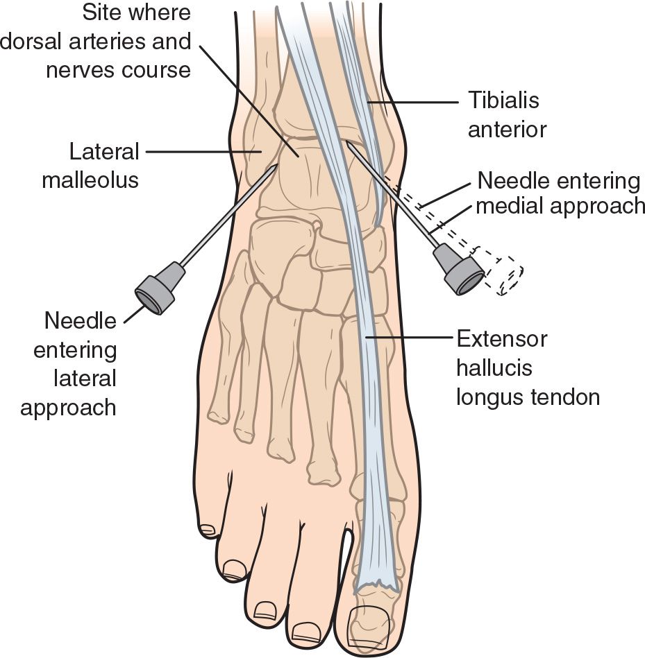
FIGURE 57.1 Arthrocentesis of the ankle. (Modified from Simon RR, Brenner BE. Emergency Techniques and Procedures. 4th ed. Philadelphia, PA: Lippincott Williams & Wilkins; 2002:246, with permission.)
Related posts:
Stay updated, free articles. Join our Telegram channel

Full access? Get Clinical Tree


