Fig.
63.1
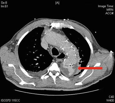
Fig.
63.2
Questions
- 1.
What is seen in the images?
- 2.
How is the above complication classified?
- 3.
What are the treatment options?
- 4.
What is the recommended surveillance to detect this complication?
Answers
- 1.
Figure 63.1 is a triple phase computed tomographic image (CT) showing a type III endoleak in the descending thoracic aorta originating from the stent graft. Figure 63.2 is also a triple phase CT image of the same patient showing the extension of the type III endoleak proximally. This patient underwent a thoracic endovascular aortic repair (TEVAR) procedure for urgent repair of acute type B thoracic aortic dissection with ongoing chest pain and rapid aneurysmal expansion. TEVAR is the procedure of choice in patients with complicated type B dissections [1]. The CT images are consistent with an endoleak. Endoleak is defined as blood collection outside the stent graft but within the aneurysm sac. This is a known complication following TEVAR. According to reported data, it occurs in about 5–20% of cases [1].
- 2.
Get Clinical Tree app for offline access
The most widely used method classifies endoleaks depending on the mechanism of formation of the endoleak [2]:
- (a)
Type I endoleak: Proximal or distal reperfusion of the aneurysmal sac. This occurs due to malapposition of the stent graft to the aortic wall. This is an early complication following TEVAR and needs urgent intervention. This is considered to be a form of treatment failure.Related posts:

Stay updated, free articles. Join our Telegram channel

Full access? Get Clinical Tree


- (a)


