Fig. 6.1
Critical pathway for the organ donor (Reprinted with permission from the United Network for Organ Sharing, Richmond, VA, 2013)
Treatment of hemodynamic instability in brain-dead multiorgan donors can be complicated by many factors unique to this patient population. In the setting of brain death, bradycardia is resistant to atropine thus beta-agonists such as epinephrine or isoproterenol should be readily available [11]. Use of vasopressors and inotropes in multiorgan donors can affect organ viability, and this patient population requires particularly careful titration of these agents. Following brain death, there is loss of catecholamine stores. Use of exogenous catecholamines can be beneficial in maintaining perfusion pressure and inotropy while helping to avoid overuse of fluids in resuscitation which can result in volume overload and negative impacts on graft function. However, high-dose dopamine and norepinephrine use has been associated with poor graft outcomes in many studies [16]. The negative effects of exogenous catecholamines in multiorgan donors is related to increases in myocardial oxygen demand, further catecholamine depletion of the myocardium, and decreased blood flow to the kidneys and liver [12].
Treating Endocrine Dysfunction
Hormonal abnormalities are common after brain-death, with diabetes insipidus being one of the most commonly encountered endocrine aberrations [15]. Hormone replacement, including the use of thyroid hormone, corticosteroids, arginine vasopressin, and/or insulin, has been shown to have beneficial effects on hemodynamic stability of the donor and organ viability [18–20]. Studies investigating the use of combined hormone therapy, with use of thyroid hormone replacement, corticosteroids, and vasopressin together, have demonstrated improvements in graft function when compared to the use of individual hormones alone [17].
Arginine Vasopressin
Following brain death, dysfunction of the pituitary and hypothalamus frequently occurs, resulting in lack of secretion of antidiuretic hormone (ADH) and neurogenic diabetes insipidus (DI) [12, 17]. If not aggressively treated, the subsequent high urine output can cause severe hypovolemia, hyperosmolality, and electrolyte disturbances, including hypernatremia, hypokalemia, hypermagnesemia, and hypocalcemia. Generally, a hypotonic solution is used with the goal of replacing urine output and baseline maintenance fluid requirements. Electrolytes should be monitored regularly, at least every 4–6 h, with goals of serum sodium less than 155 mEq/l and serum potassium greater than 3.5 mEq/l [12].
Arginine vasopressin (AVP) is commonly used in cases of severe DI in brain-dead patients, and studies have demonstrated improved hemodynamics and reduced inotropic requirements when such patients are treated with AVP [21]. Pennefather et al. [21] compared hemodynamic parameters in brain-dead multiorgan donors treated with low dose AVP infusion (dose of 300 mU/kg/min) versus saline infusion. This study determined that the use of AVP was associated with reduced dopamine requirements and improved blood pressure without worsening of hemodynamic parameters or graft function. Pennefather et al. [21] concluded that use of a vasoconstrictor, such as AVP, in brain-dead patients is beneficial given the loss of vasomotor tone often found in such patients and that, frequently, inotropic agents are inappropriately used in this setting. It is recommended that AVP be given as a low dose infusion in order to avoid the potential for end organ damage with high dose vasoconstrictor use.
Desmopressin , a synthetic vasopressin analogue commonly used to treat DI, has an antidiuretic-to-pressor ratio of 2000 to 4000:1 compared with a ratio of 1:1 seen with AVP. Studies comparing treatment of multiorgan donors with desmopressin versus AVP have demonstrated that both are equally effective in treating DI symptoms, such as high urine output, and resulted in equivalent kidney graft outcomes. Thus, both desmopressin and AVP are effective in treating DI in the brain-dead patient; however desmopressin is less effective in elevating system blood pressure in such patients given its profile as a weak vasopressor when compared to AVP [17].
Thyroid Hormone Replacement
Use of triiodothyronine (T3) and thyroxine (T4) replacement in brain-dead patients has been found to be associated with improvement of metabolic acidosis and hemodynamic instability in brain-dead patients while lowering requirements for both bicarbonate and inotropic agents [15]. It has been postulated that brain death results in decreased levels of T4 and T3, hormones necessary for storage of energy in the myocardium, ultimately resulting in hemodynamic instability [14, 17].
Replacement of thyroid hormone does, however, remain controversial in this patient population. Goarin et al. [22] postulated that T3 is low in brain-dead patients and that this euthyroid sick syndrome must be a component of myocardial dysfunction seen in this patient population. However, when comparing patients given a placebo versus T3, no difference was found in hemodynamic parameters between groups or in echocardiography interpretation. Other studies have demonstrated similar results and found little correlation between thyroid hormone levels and degree of hemodynamic instability.
In contrast, other studies have demonstrated hemodynamic improvement in patients treated with thyroid hormone. In one study by Salim et al. [14], hemodynamically unstable brain-dead patients showed a statistically significant decrease in vasopressor requirements after treatment with T4. Such studies indicate that use of thyroid hormone replacement improves energy metabolism in the myocardium thus reducing acidosis and improving cardiac function.
Given that T4 is a prohormone which is converted to biologically active T3, T3 is generally considered first line for thyroid hormone replacement in this patient population [17]. Given the continued controversies regarding the efficacy of thyroid hormone replacement in brain-dead multiorgan donors, there remains much variability between institutions in its use for this indication and further research is indicated.
Corticosteroids
At the time of brain death, a multitude of proinflammatory cytokines are released, resulting in hemodynamic instability. It has been postulated that corticosteorid use attenuates the release of these proinflammatory mediators, benefitting both the donor throughout the organ procurement process and the recipient after subsequent organ transplantation. Additionally, the stress response associated with brain death can result in adrenal insufficiency, providing another proposed mechanism for benefit of corticosteroids in brain-dead patients [23].
Controversy exists regarding the degree of adrenal suppression found in brain-dead multiorgan donors. Dimopoulou et al. [24] found that adrenal cortisol secretion was impaired after stimulation in brain-dead patients; however, other studies have demonstrated normal adrenal function in brain-dead patients during the period surrounding organ procurement. Several studies have failed to demonstrate an abrupt decline in cortisol levels, indicating that perioperative steroid replacement may not be advantageous in brain-dead multiorgan donors 16].
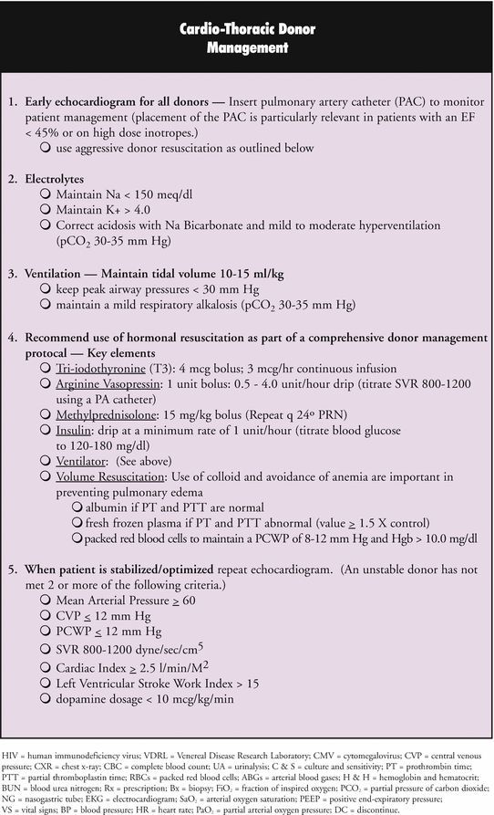
Although some studies question the need for corticosteroids in all organ donors, many institutions and transplantation programs include corticosteroids in the perioperative care of multiorgan donors. Currently, UNOS recommends a bolus dose of methylprednisolone in brain-dead multiorgan donors in preparation for organ procurement and has included this in the Critical Pathway for cardio-thoracic donors (see Fig. 6.1).
Insulin
Hyperglycemia is a common manifestation of brain death, and many studies have looked to determine if the cause is pancreatic dysfunction secondary to brain death. Overall, evidence shows that endocrine pancreatic function remains effective in most patients after brain death and that hyperglycemia is, most likely, secondary to peripheral insulin resistance [2, 25]. Exogenous catecholamine administration and physiologic stress place brain-dead patients at particular risk for development of insulin resistance [17]. Regardless of the cause, treatment of hyperglycemia is necessary in brain-dead patients as hyperglycemia has been shown to result in osmotic diuresis, electrolyte abnormalities, and worsening of graft function. Lung and heart grafts appear to be particularly susceptible to the negative effects of hyperglycemia [2, 18].
Anesthetic Agents During Organ Procurement
After brain death spinal reflexes remain intact, and thus spontaneous movement is possible in brain-dead patients. During organ procurement it is recommended that neuromuscular blocking agents be given in order to prevent movement and provide optimal surgical conditions [2, 12]. It is commonly recommended that brain-dead donors do not require anesthesia during organ procurement. However, the fact that brain-dead multiorgan donors demonstrate increased release of catecholamines in response to painful stimuli during organ procurement has prompted several studies to examine the role of opioids in such patients. A study by Fitzgerald et al. [26] found that a dose of 7 μg/kg of fentanyl did not suppress this catecholamine discharge or attenuate the hemodynamic response to surgical stimulation when compared to placebo during organ procurement in brain-dead donors.
Related posts:
 Preoperative Evaluation and Preparation for Lung Transplantation
General Anesthesia for the Patient with End-Stage Liver Disease and Post Liver Transplantation
Preoperative Evaluation and Preparation for Lung Transplantation
General Anesthesia for the Patient with End-Stage Liver Disease and Post Liver Transplantation
 Anesthetic Management of Patients Undergoing Pancreas Transplantation
 Acute Liver Failure: Perioperative Management
Postoperative Care of Heart Transplant Patients
Anesthesia for Composite Tissue Allografts
Acute Liver Failure: Perioperative Management
Postoperative Care of Heart Transplant Patients
Anesthesia for Composite Tissue Allografts
Stay updated, free articles. Join our Telegram channel

Full access? Get Clinical Tree


