and Richard A. Jaffe2
(1)
David Geffen School of Medicine at UCLA, Los Angeles, California, USA
(2)
Stanford University School of Medicine, Stanford, California, USA
Keywords
FiresFire triadPreventionIntroduction
Oh no! Not again!! Not another operating room fire!!! How does this keep happening? Here we are, a very sophisticated, high technology country and we still can’t seem to prevent fires in the operating room. Why is that? What are we doing wrong? While there has been a general awareness of operating room fires for many years, with many publications, especially in the journal Anesthesia and Analgesia, of fires occurring under various circumstances, operating room fires continue to occur. Because of this, the American Society of Anesthesiologists created a Task Force on Operating Room Fires, and their report appeared in Anesthesiology [1]. Included in the article is an Operating Room Fire Algorithm (Fig. 16.1), which, like most algorithms created by committee, is complex and difficult to remember in detail. To be useful in an emergency, it would need to be posted in a prominent location in each operating room, and this is what the ASA recommends. In addition to the ASA publication, the Anesthesia Patient Safety Foundation has prepared a short video on the prevention and management of operating room fires that is available for free by contacting the APSF (www.APSF.org). The APSF has also developed a simpler algorithm for fire prevention (Fig 16.2). The message from both sources is the same, and will be the essence of this chapter. Two case examples are provided to illustrate the types of issues that arise and result in an operating room fire.
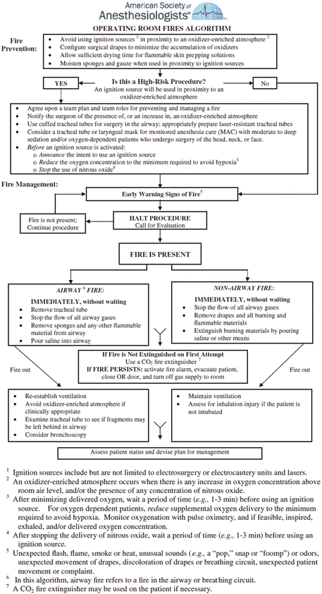

Fig. 16.1
Operating room fire algorithm recommended by the American Society of Anesthesiologists (ASA). The ASA also recommends that this algorithm be posted in every operating room. From: Ref. [1]. Copyright © by the American Society of Anesthesiologists, reprinted with permission
Fig. 16.2
Fire algorithm recommended by the Anesthesia Patient Safety Foundation (APSF) . Reprinted with permission
Case One
The patient is an 84-year-old woman who was seen by her dermatologist because of a lesion on her nose. The dermatologist thought that the lesion was cancer so he referred her to a surgeon. Her history was significant for hypertension for which she took Norvasc 10 mg bid. In addition, she had a past history of congestive heart failure, although she was asymptomatic and her lungs were clear at this time. She had a history or coronary artery disease , and stents had been placed several years earlier. She had no history of chest pain or shortness of breath while walking or climbing a flight of stairs. She had a history of spinal stenosis and spondylolisthesis for which she took analgesics occasionally. Finally, she had a very recent right rotator cuff tear following a fall, for which she took analgesics (oxycodone) especially at bedtime.
After a complete workup, the surgeon admitted her to an outpatient facility for removal of the lesion on the nose. The plan was to excise it with electrocautery under monitored anesthesia care (MAC) . The anesthesia provider applied all of the monitors and then attached a nasal cannula through which he delivered oxygen at 5 L/min. Just prior to the start of surgery, he administered midazolam 2 mg and fentanyl 100 mg to the patient. Shortly after the start of surgery, the patient complained of discomfort, so she was given both drugs in the same dose again. The lesion on the nose was excised with a cold knife and the bleeding controlled with the electrosurgical cautery. The tissue was sent to a pathologist who indicated that the margins were not clear, and recommended a wider excision. A second specimen was taken and the bleeding again controlled using the cautery. Shortly thereafter the surgeon noted flame under the drape. He removed the drape immediately and noted that the nasal cannula and oxygen delivery hose were burning. The anesthesia provider turned the oxygen off and removed the nasal cannula. In the process of removing the flaming drapes, the oxygen hose hit her right shoulder causing an additional burn.
Postoperatively she had second degree burns of her nose, face, neck and shoulder. The burns were cleansed and coated with SilvadeneTM . She was admitted to hospital and over the next week her burns started to heal. Over the subsequent weeks she developed stenosis of her nasal passages, which required surgical correction. Upon discharge from hospital a month after the injury, she continued to complain of difficulty breathing through her nose and pain in her face and mouth. After 1 year, her facial appearance became more tolerable for her.
Case Two
The patient is a 74-year-old woman who was diagnosed with bilateral ptosis of the eyelids that was compromising her vision, and was scheduled by a plastic surgeon for a bilateral blepharoplasty and possible brow elevation. Her past history included a myocardial infarction 12 years earlier that was treated with stent placement. She had a history of hypertension, which was treated with LosartanTM , and HyzaarTM , moderate aortic regurgitation that was not treated, and hypercholesterolemia that was treated with atorvastatin (LipitorTM). She was seen by her cardiologist preoperatively and he concluded that she was only a mild risk for postoperative cardiothoracic complications as evidenced by her absence of any chest pain, shortness of breath or other symptoms, a recent negative stress echocardiogram, an unchanged EKG, and that she functioned normally at an oxygen saturation of 94 % breathing room air.
On the morning of surgery in the ambulatory surgery center, the surgeon announced that he would be using electrosurgical cautery to control bleeding, and that he needed the patient reasonably awake at the end of the blepharoplasty, so he could have the patient sit up and he could determine if any additional muscle resection was needed to correct the ptosis. The plan was to use local anesthesia on the eyelids and MAC with light sedation. The anesthesia provider insisted on using a mask instead of a nasal cannula for better oxygen provision. She trimmed the edges of the transport mask so they would not be in the surgical field. She turned the oxygen flow to 4–5 L/min. For sedation she gave the patient diphenhydramine 25 mg and hydromorphone 1 mg i/v.
The surgeon completed the operation on the right eyelid in about 45 min, and then started on the left eyelid. Shortly thereafter the surgeon noted a spark and then flames in the drape. He pulled off the drape and saw fire in the mask and oxygen tubing. The anesthesia provider shut off the oxygen flow and the scrub tech poured sterile water on the patient’s face. A plastic surgeon was called to the OR and he diagnosed second and third degree burns of the face, nose eyelids and neck. Because of concern about an airway burn, the anesthesia provider administered a small dose of propofol (80 mg) and rocuronium (40 mg) and intubated the trachea. A pulmonologist came to the OR and did a bronchoscopy, which showed no injury to the larynx, trachea or bronchi. The wounds were coated with ointment and she was taken to the hospital ICU. Her trachea was extubated the next day and 4 days later she was transferred to a burn center. She remained at the burn center for several weeks, and had multiple surgical procedures to correct scar tissue formation. She remained blind in her left eye.
Related posts:


Stay updated, free articles. Join our Telegram channel

Full access? Get Clinical Tree


